POTD: High-Pressure Injection Injury

High-Pressure Injection Injury

·      Patients present with seemingly innocuous findings after high-pressure injection injury

·      Their condition often rapidly deteriorate

·      Substances can be paint, paint stripper, grease, oil, water or air.

·      This is a surgical emergency and early consultation is critical for surgical decompression and debridement

·      Less viscous substances can penetrate deeper with less pressure, leading to worsened outcomes, even if initially the wound may appear benign on the exterior, and even if the patient’s pain is initially minimal

·      Paint and paint thinners produce a large and early inflammatory response leading to ischemia and tissue death and the rate of associated amputation is high.

·      Initial emergency department management:

o   pain control, radiographs (look for free air), elevation, splinting, IV antibiotics, tdap, emergent hand specialist consultation

o   These injuries are not high-risk injuries for tetanus, and prophylaxis, even if indicated, therefore tdap should not delay other steps in management.

o   In fact, none of the emergency department interventions, (besides pain control), is as important as recognition of the potential severity of the injury and early consultation with a hand specialist

o   There is no amount of cleansing this wound in the ED that is recommended because the penetration is deep and this patient needs to go to the OR.

·      It is interesting to note that although digital blocks are excellent tools to relieve pain and provide anesthesia, they are not recommended in high-pressure injection injury as one of our major concerns is compartment syndrome.

o   Digital blocks can lead to an increase in compartment pressure and worsen injury/tissue ischemia. Systemic pain control is recommended.

The below picture is of a hand in the OR, you can see the initial presentation appears someone benign and once the hand is opened up, you see a lot of tissue necrosis.

potd high pressure.jpg

Below pictures show benign physical exam findings and some free air on xray

potd finger.png

Sources: Tintinalli, Rosen's Emergency Medicine, uptodate, Peer IX, ortho blog for photos: http://www.cmcedmasters.com/ortho-blog/high-pressure-injection-injuries

 · 

POTD Trauma Tuesday: Name that fracture!

A 36-year-old man presents by ambulance following a motorcycle crash. He told the EMTs that he lost control and fell sideways, bracing his fall with his outstretched right hand. His R arm looks deformed but is neurovascularly intact. An xray is obtained.

xray potd.png


What’s the name of this one again?!

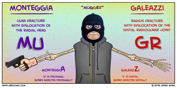
Galeazzi fracture! Named after an Italian surgeon from Milan.

What is it? a fracture-dislocation of the distal third of the radius associated with dislocation-subluxation of the distal radial ulnar joint, or DRUJ.

Why do we care? With Galeazzi fractures, there is a high risk of malunion, loss of function, infection, and chronic pain in adult patients. For this reason, surgical management with internal fixation and possible open reduction is required. The repair should occur promptly, so the emergency physician or another clinician should contact the orthopedic consultant emergently to coordinate care.

What about in peds? Emergent orthopedic consultation is still required but interestingly, in children, some Galeazzi fractures are treated conservatively with closed reduction by an orthopedic surgeon. Disruption of the DRUJ can be subtle, so a high suspicion should be maintained when a patient presents with a fracture of the distal third of the radius.

Pearls of the Peal:

* Look for fracture-dislocation of the distal radius and ulna after a fall onto an outstretched arm. This injury can’t be missed: it requires immediate orthopedic involvement.

* Skin tenting associated with the Galeazzi fracture-dislocation puts the patient at risk for skin necrosis and conversion to an open fracture.

Wasn’t there some way to remember this compared to other one?? Why, yes! See below:


mugger.png

Sources:

Comic: Medcomic.com

Xray and clinical information: PEER IX

 · 

POTD: Straight leg test. A leg up on clinical testing!

A little background:

Lumbar disc herniation is the most common cause of lumbar radiculopathy, or sciatica, a shooting or burning pain from the low back radiating down the posterior leg distal to the knee.

Two tests used to evaluate these symptoms are

The straight leg raise.

·       The straight leg raise test is highly sensitive but not very specific for disc herniation.

·       This is performed by lifting the leg affected by the radiating pain.

·       The patient lies supine with one leg either straight or flexed at the knee with the sole of the foot flat on the stretcher.

·       The examiner then raises the affected leg up, extended, to 30 to 70 degrees.

·       Reproduction of low back pain that radiates down the posterior affected leg at least past the knee is considered a positive result. Not just pain to the lower back, which is a common misconception.

·       The SLR test can also be performed with the patient in a sitting position, by stretching the sciatic nerve by extending the knee; the test is positive if pain radiates to below the knee.


The crossed straight leg raise.

·       It is highly specific (90%) for disc herniation

·       You perform the same test as the straight leg but on the unaffected leg.

·       A positive test: reproducing both the back pain + the radiation down the affected leg.

Sources: Peer IX, Tintinelli’s, Dr. Sergey Motov, Uptodate

 ·