VOTW: Pediatric Skull Fracture

This week’s VOTW is brought to you by the UST~

A 9 month old female infant was brought into the Pediatric ED two days after a fall from a high chair. The infant vomited once after the fall but was otherwise acting normally since then. The patient was brought to the ED 48hrs after the fall for a boggy left parietal scalp hematoma. The patient had a normal physical exam apart from the hematoma.  A POCUS was performed which showed...

Clip 1 shows an oblique disruption in the cortex of the skull, indicative of a fracture. The bones have an “overlapping” appearance. A hypoechoic hematoma is present overlying the fracture.

Image 1 shows the same fracture with relevant structures labeled.

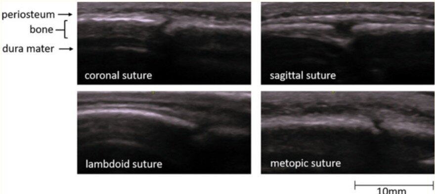
Image 2 shows a cortical disruption in the skull of the same patient, but this one is a cranial suture

Sutures and fractures look the same! How do I differentiate them?

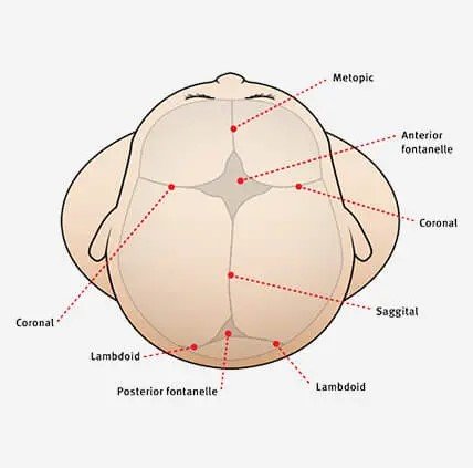
  • A suture can be followed all the way to a fontanelle.

  • Sutures are present symmetrically - scan the contralateral side if unsure

  • Fractures may appear irregular, jagged or displaced.

  • Sutures generally have an “end-to-end appearance” (image 2)- the cortex stops, there is a small space, and then restarts.

  • A fracture is likely to have an overlying hematoma.

Image 3. More examples of sutures

Image 4. A review of the anatomy of sutures and fontanelles

How to perform the study

  1. have a parent or assistant stabilize the child’s head, especially if they are squirmy

  2. use a linear high frequency probe and a lot of gel, especially if there is hair

  3. warm up the gel (put the gel bottle in your backpocket) which might make it less uncomfortable for the patient

  4. scan the area of swelling in two orthogonal planes and look for disruptions in the cortex

  5. scan the area around the hematoma as well- the fracture may not be directly under the hematoma

Clinical Decision Making

There is limited data on the use of POCUS for diagnosing pediatric skull fractures.

  •  When performed by EM Physicians, POCUS for skull fractures has sensitivities ranging from 67% - 100% and specificity of 85% - 100% (1)

  •  The presence of a skull fracture increases the likelihood of intracranial injury by four-fold (2)

POCUS for pediatric skull fractures might be most useful in the borderline case- for example a child who has an occipital/parietal/temporal scalp hematoma but otherwise looks great in the ED. Using PECARN you decide that you would rather observe this patient than subjecting the patient to radiation +/- sedation. If you decide to perform a POCUS, the absence of a skull fracture might be reassuring to you (and the family) and support your shared decision to observe the patient. The presence of a skull fracture might raise your concern for intracranial injury and change your decision about imaging. 

For a patient with a high pre-test probabiltiy for underlying pathology a negative POCUS should not be used a rule out test.

It might also be useful seeing a depressed or complex skull fracture as this may expedite imaging and specialist consultation.

More research is needed to define the role of POCUS in clinical decision making and how we might be able to integrate it with clinical decision rules like PECARN.

Happy Thanksgiving!

Your Sono Team

  1. Alexandridis G, Verschuuren EW, Rosendaal AV, Kanhai DA. Evidence base for point-of-care ultrasound (POCUS) for diagnosis of skull fractures in children: a systematic review and meta-analysis. Emerg Med J. 2022 Jan;39(1):30-36. doi: 10.1136/emermed-2020-209887. Epub 2020 Dec 3. PMID: 33273039; PMCID: PMC8717482.

  2. Kuppermann N, Holmes JF, Dayan PS, et al.. Identification of children at very low risk of clinically-important brain injuries after head trauma: a prospective cohort study


VOTW: Intussusception

Today’s VOTW is brought to you by Dr. Fagan, Dr. Davitt and Dr. Lat!

A 2 year old male presented with abdominal pain and vomiting x1 day as well as cough and nasal congestion x2 days. On exam, he was clutching his abdomen in discomfort. A POCUS was performed which showed…

Clip 1 shows an abdominal ultrasound in the RUQ showing the classic “target sign” measureing 3.5cm, concerning for intussusception. You can visualize a smaller circular structure within a larger circular structure representing a part of bowel telescoping into the next part of the bowel. In the center, there are small circular hypoechoic lymph nodes surrounded by echogenic mesenteric fat that serves as the leading point of the intussusception.

Image 1. Target or donut sign

Ultrasound is the test of choice for intussusception and several studies have shown high sensitivity (94%) and specificity (99%) when POCUS is performed by PEM physicians (2).

POCUS for Intussusception

  • Most commonly occurs at the ileo-cecal junction and most commonly found in the right lower or right upper quadrant

  • Look for a target sign or donut sign (in transverse view, see above) or sandwich or pseudokidney sign (in longitudinal view, see below)

  • Diameter > 2cm (remember in-✌-ssusception)

    • May see mesenteric fat and lymph nodes in the center

Image 2. Pseudokidney or sandwhich sign

How to perform the study            

  • Use warm gel, have parents help distract, scan on parent's lap!

  • Use the linear probe

  • Picture frame pattern- start in the RLQ w/ probe marker to pts R scan towards the RUQ, then turn the probe w/ marker to pts head and scan towards the LUQ, then turn the probe w/ marker to pts R and scan down to the LLQ

  • Lawnmower pattern- start in the RLQ and lawnmower the entire abdomen scanning up and down from right to left with the probe marker to the pts R

  • Measure the diameter of the intussusseption if found

Image 3. Picture frame pattern

Back to the patient

Surgery was consulted, the patient underwent an air enema with resolution of the intussusseption. The patient was then discharged home.

References:

  1. Lin-Martore. PEM POCUS Series: Intussusception. https://www.aliem.com/pem-pocus-series-intussusception/

  2. Lin-Martore M, Kornblith AE, Kohn MA, Gottlieb M. Diagnostic Accuracy of Point-of-Care Ultrasound for Intussusception in Children Presenting to the Emergency Department: A Systematic Review and Meta-analysis. West J Emerg Med. 2020 Jul 2;21(4):1008-1016. doi: 10.5811/westjem.2020.4.46241. PMID: 32726276; PMCID: PMC7390574.


POTD: Neonatal Resuscitation

We’ll be going over a few high yield topics pertaining to NALS today. 

It’s 7:30 AM, and you’ve just unwrapped your BEC sandwich and taken your first sip of coffee. You’re settling into the morning getting ready for your 12 hour peds shift… until the phone rings, and you get a note: 

“Mother 38w delivered her baby at home 30 minutes ago. Baby is having labored breathing, and is bradycardic. EMS will be here in 2 minutes.”

Take a deep breath. First, remember the basics. If you’re in a facility that has Peds/NICU, call them immediately. Call respiratory. Call pharmacy. Call Hector. Use the resources available to you. 

The set up.

Get the warmer and set it to 25 C

  • Avoid hypothermia in these patients. The goal is > 36.5-37.5C

Grab the Broselow tape so that it’s available for immediate use.
Get the backboard.
Grab the code cart, zoll
Get a towel to warm and dry the baby.
Get your airway equipment ready:

  • Suction x 2, plugged in, ready to go

  • Oxygen: grab the neonatal BVM and plug it into the oxygen port

  • Airway equipment: have both DL/VL equipment,

    • LMA size 1

    • Pre-loaded tubes

      • 2.5 and 3.0 uncuffed tubes

    • Blades: 0 and 1

    • EtCO2

Access: IO gun + pink needles ready for use; umbilical vein catheters (future POTD)

Grab your PALS card or open up your PediStat app
Ultrasound

Assess the patient.

Pediatric assessment triangle:

  • Appearance – crying? Good tone? Tracking?

  • Breathing – nasal flaring? Stridor? Grunting? Head bobbing?

  • Circulation – Pallor? Cyanosis? Mottling?

Off the bat, there are two numbers you need to remember:
HR < 100→ initiate positive pressure ventilation (PPV)
HR < 60→ initiate CPR / epinephrine if this is sustained more than 30 seconds despite adequate ventilation.

  • NOTE: Bradycardia is almost always related to hypoxia, so atropine isn’t routinely indicated for these patients.

Remember, the most important part of neonatal resuscitation is positive pressure ventilation.


PPV.

If the patient is spontaneously breathing but labored, you can place them on CPAP.
Remember, the targeted SpO2 after birth is much lower for neonates, so see the box below. You’re more interested in ventilating than the oxygenation.
For gasping / apneic / HR < 100 patients, initiate PPV. You can use 5 on the PEEP valve.

  • Rate: 40-60 breaths / minute

MR SOPA mnemonic for ventilation tips:

  • Mask, right size

  • Reposition airway

  • Suctioning nares

  • Open mouth

  • Pressure increase to PEEP to ~5

  • Advanced airway: ETT / LMA

BGM.

They also have lower BGMs. Hypoglycemia for neonates is < 30 for a patient < 24 hours old. It’s recommended to give D10 bolus 2ml/kg if the patient is hypoglycemic.

You can give glucagon IM too: 0.03mg/kg max 1mg

CPR.

It’s recommended to secure an airway (supraglottic or ETT) prior to doing compressions) since most these codes are usually due to respiratory events.
The ideal ratio is3 compressions:1 breath

  • Goal is 90 compressions: 30 breaths in one minute

2 thumb compression technique (*preferred) or 2 finger technique
Pulse checks q1 min
Depth: ⅓ chest diameter

Epinephrine.

IV dosing: 0.01mg/kg q3-5min
ETT dosing: You can give epinephrine through the ETT too if you don’t have access yet! AHA recommends a larger dose 0.1mg/kg of 1:1000 ETT

  • Max dose is 10mg, and follow it with a saline flush

I highly recommend reviewing the following flowchart linked.

I hope this was a good refresher on some of the most important concepts. I would love to learn other tips that others have in managing these stressful situations!

References:

https://cpr.heart.org/en/resuscitation-science/cpr-and-ecc-guidelines/neonatal-resuscitation 

https://emergencymedicinecases.com/neonatal-resuscitation/