To Ponder Puncturing the Peritoneum?

When and when not to perform paracentesis? Think of the indications for emergent paracentesis in a similar fashion to emergent thoracostomy.  Indications:

  • Relief of respiratory distress caused by massive ascites

  • Diagnostic for infection ie. suspected spontaneous bacterial peritonitis

Contraindications?

  • Overlying cellullitis

  • Vasculature or bowel obstructing desired site

  • Loculated fluid (concern this may be oncologic)

  • Significant coagulopathy INR < 8 platelets >20

2 minute EMRAP video outlining the procedure- https://youtu.be/9npNQM8ANds

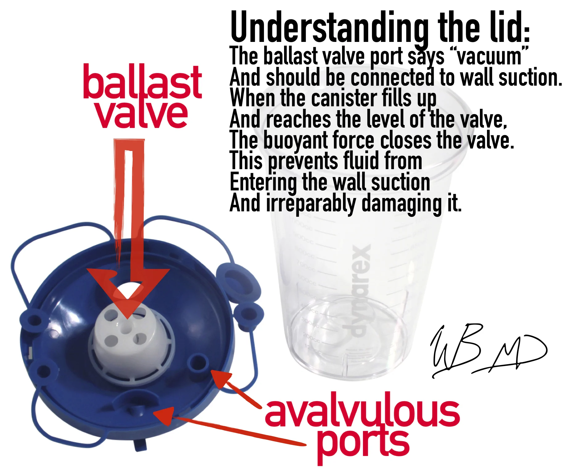
Blog post explaining in-series suction canisters-  http://mmcedrco.w02.wh-2.com/EMBlog/suction-cannisters-in-series/


 · 

Pulse Cooximeter: how to use it

On my previous shift, we had 2 patients with a “gas” exposure at their apartment. 

So the clinical question arose: How can we rapidly screen patients with “gas” exposure? 

First, any abnormal vitals raise a red flag and automatically take us off the rapid discharge pathway. These patients need appropriate triage to (

likely

) north side. 

In any patient who has been exposed to a fire or there is concern for carbon monoxide exposure, we have the Masimo pulse-cooximeter. 

It is located in the south side charge nurse station. 

To utilize this device: hold down the power button while the pulse sensor is on the patients finger.

masimo_co-oximetry.jpg

After pressing the display button, it should look as below:

Please note that there is a continuous SpCO and SpMet in green on the left and right of the display, respectively. 

If results in an asymptomatic patient with a low risk exposure are normal, they can be safely discharged without further testing. However, in a symptomatic patient with a normal pulse-cooximetry, they should be further screened with blood gas cooximetry. Furthermore, a

ny abnormal value of %SpCO>5% should be repeated with a blood-cooximetry.

Smokers may have a baseline CO-Hgb of 5-6%, and may require confirmatory testing with blood-cooximetry through our blood gas lab. 

In short, if patient has a %SpCO <5% and is asymptomatic they may be safely discharged. This also requires a normal %SpO2 because %SpO2<85% decreases the accuracy of the Masimo pulse co-ox, as per the literature posted on their own product page. 

Final summary of this POD:

we have a pulse-cooximeter.

Utilize it for rapid screening and for reassurance of low risk patients. 

Please clean the finger sensor between patients with a purple wipe

As always: feedback both negative and positive IS STRONGLY ENCOURAGED. 

TR,

Wells 

 · 

Suction cannisters in series

Today during my TR shift, CY asked me for demonstration of how to connect suction canisters in series for large volume paracentesis.

Before we go into the actual technique, lets take a second to understand how the suction canister functions. This is not our exact model of suction canister lid, but is functionally the same. See my infographic below:

suction-kanister-explained.jpg

The goal: to connect 3 of the vacuum canisters together so that when one fills, the fluid then starts draining into the next. Once the second fills it will drain into third and automatically stop.

With this technique you can perform your paracentesis, start drainage, and then safely walk away. Please instruct your patient not to move whilst drainage is in action.

So connect one suction canister with vacuum tubes as you normally would: vacuum to wall suction; the next vacuum tube should go from either of the avalvulous ports to the next avalvulous port. Critical point: the only port that needs to be connected to the ballast valve port is the port that plugs into wall suction. All subsequent ports should be connected in series with the avalvulous ports.

The final attachment is the avalvulous port to christmas tree adapter, which is included in our thora/paracentesis kit.

During what I believe was my intern year, I had shown Strayer this technique, and much to my chagrin, he tweeted out a post about it. See below:

strayers_ccannister-post-1.jpg
suction-kanister-explained.jpg

The benefits of utilizing this technique for large volume paracentesis are twofold: (1) increased safety and (2) utilization of resources (in this case physician time). Safety is increased because this method does not utilize sharp needles to transfer fluid to a vacuum flask; thereby, it reduces the risk of needle stick injury (especially important for the population of patients that need ED paracenteses). Utilization of resources is improved because a physician does not need to remain at bedside or repeatedly check the progress of evacuation, assessing for need to switch to the next vacuum flask.

For large volume paracentesis, I recommend only connecting a max of 4 of our suction canisters in series. I typically do 3, but 4 approaches that max of 5L that current thought suggests may cause hemodynamically significant fluid shifts.

 ·