POTD: Chest Tube/Pig Tail Drain Suturing

There are several different techniques, so I wanted to illustrate a few of them. The common theme is start from above or below the chest tube and throw your first knot to the skin:

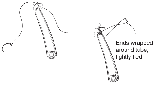
1) Wrapping technique (this is my go-to technique)

- begin by throwing your first knot into the skin above the tube, then wrap the suture material around the base of tube (at the level of the tube insertion site)multiple times and then tie into place

- this can be repeated from below the tube for extra security

- video: https://www.youtube.com/watch?v=v2y-g0RAImw

mint_c044f006.gif

2) The “ps and qs” technique (here called the easy L)

- essentially throw your first stitch in the skin and then perform 3 “p” stitches or 3 “q” stitches and vice versa

- finish by performing hand ties

- video: https://www.youtube.com/watch?v=Qsq1fPxYNrQ

3) The “big S” technique

- similar to a nautical knot, called the clove hitch

- essentially form an “S” shape underneath the chest tube (after you throw your first stitch)

- video: https://youtu.be/4lkyq7U6fpg

Other techniques include purse string sutures or horizontal mattress sutures.

 · 

Targeted Temperature Management

Targeted Temperature Management

What is it: the purposeful cooling of a patient post-cardiac arrest. Target of 32°C to 34°C (Some studies say 36, but debatable and prevent any hyperthermia) for at least 24 hours. 

Why: To improve the chance of survival and neurologic recovery, international guidelines recommend use of targeted temperature management (TTM), together with urgent coronary angiography and percutaneous coronary intervention when appropriate

Who: 

  • Post cardiac arrest (any cause but most evidence supports from VF/VT shockable causes of cardiac arrest)

  • ROSC < 30 mins from team arrival

  • Time < 6 hours from ROSC

  • Patient is comatose, GCS <8 (this is try and improve neurological outcome, so someone who is neurologically intact does NOT need TTM)

  • MAP >= 65mmHg

  • depends on your hospital protocol

When: Initiate within 6 hours of ROSC and maintain for 24 hours

How: 

  • cold IVF at 2-3 mL/kg stat

  • cooling vest and cooling machine

  • sedation and paralysis

Thumbnail_TemperatureManagement_AC.jpg

Complications:

Shivering, electrolyte abnormalities, cold diuresis, infection. 

IJCCM_2015_19_9_537_164806_t1.jpg

So, for post cardiac arrest patients with depressed neurological function - Keep this in mind, but consult your ICUs and plan this patient's care together for best management. TTM needs an ICU level care admission. 

Happy Learning!

References:

https://jamanetwork.com/journals/jama/fullarticle/2645105

https://www.ncbi.nlm.nih.gov/pmc/articles/PMC4578199/
https://lifeinthefastlane.com/ccc/therapeutic-hypothermia-after-cardiac-arrest/

http://www.ijccm.org/article.asp?issn=0972-5229;year=2015;volume=19;issue=9;spage=537;epage=546;aulast=Saigal

 · 

Do or Di-alysis there is no try... unless you use a trialysis catheter

Ok, lets review the indications for emergent dialysis and the process of placing a dialysis catheter. Indications: -severe acidosis secondary to renal failure or unresponsive to medical therapy -toxic ingestion that is small not protein bound, such as alcohols salicylate, lithium, theophylline, valproate - symptomatic hyperkalemia - symptomatic hypernatremia -fluid overload with oliguria causing respiratory failure -uremia causing encephalopathy, pericarditis, or hemorrhage

placement:

**review your own kit to make sure you have everything you will need and to be mindful of possible extra steps for the following

  1. give anxiolysis if needed

  2. position patient and ultrasound for best place for your catheter

  3. sterilize skin

  4. prep your kit, gown, hat, mask, gloves, sterile cover for ultrasound

  5. flush lumens of your cathetern and apply lumen valves to each lumen but do not place one on the most distal luman through which your wire will pass

  6. have sterile heparin drawn up

  7. drape patient

  8. anesthetize skin

  9. find view use ultrasound guidance and advance needle into vein be sure to be drawing the syringe plunger back to aspirate for blood

  10. once in the lumen advance the needle slightly more in the middle of the lumen to prevent loosing your placement

  11. with non-dominant hand flatten need while making sure to not pull out of the vein

  12. remove syringe from the needle and advance your wire *if there is resistance ultrasound to recheck your placement

  13. advance wire while holding it securely (be sure to always have at least one hand holding the wire)

  14. remove needle

  15. load smaller dilator onto wire

  16. cut the dermis at wire insertion be sure to cutaway from wire

  17. advance dilator and push through skin with twisting motion and inline with trajectory of the wire

  18. remove dilator

  19. load second larger dilator and repeat steps 17 & 18

  20. remove dilator there will be lots of blood good job

  21. advance catheter holding close to the skin with a firm but gentle twisting motion

  22. remove wire

  23. check that all lumens draw back blood with ease

  24. flush each lumen with 1cc of heparin to prevent clotting of the catheter

  25. secure the catheter with sutures

  26. apply sterile dressing

  27. if in internal jugular of subclavian veins confirm placement with xray

 ·