Electrical Storm

storm.jpeg

Electrical Storm


Background

  • three or more episodes of sustained ventricular tachycardia, ventricular fibrillation, or appropriate ICD shocks in a 24 hour period

  • frequently, hemodynamic unstable

  • believed to mostly be due to catecholamine surge (sympathetic overdrive), but need to consider the causes below:

    • MI

    • Electrolytes

    • Acute HF

    • QT prolongation/shortening

    • Torsades

    • Brugada

    • Thyroid storm

    • Drugs

    • Sepsis

  • Presentation is broad

    • May complain of pain from ICD shocks, palpitations, syncope

    • Can present in cardiac arrest
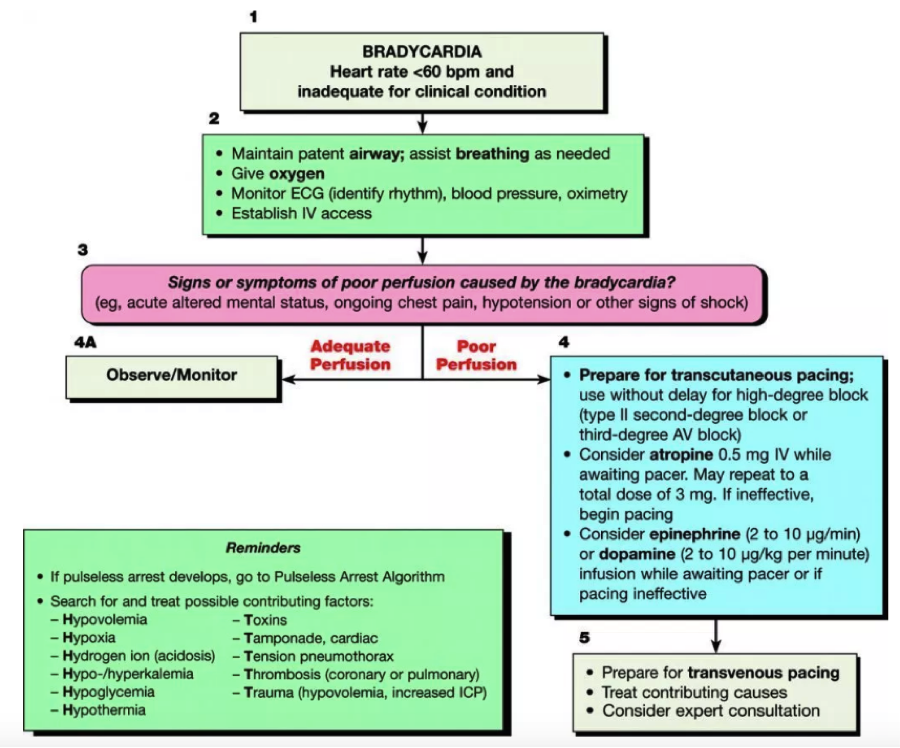
Management

  • ABCs

  • ACLS guidelines should be followed

    • Pulse (VT)==>cardiovert

    • Pulseless (VT/VF)==>defibrillate

      1. Consider dual defibrillation if VT/VF persists after 5 delivered shocks (see image below)

        1. Coordinate firing of both defibrillators at the same time

dual defib.png

  • Should you place a magnet over ICD if patient has one?

    • Remember, magnets turn of the ability to defibrillate, but don't affect pacing capability

    • If you want the patient to be shocked then do not place a magnet, unless you think you’re dealing with something other than VF/VT and thus it is shocking inappropriately (sinus tach, afib)

      1. Practice variation exists. Some will place the magnet, especially if the pt is stable, to spare pt more anxiety/pain contributing to the storm

  • Start them on an anti-arrhythmic

    • Amiodarone 300mgè150mg IV //

    • Procainamide 10 mg/kg IV over 20 min //

    • Lidocane 1-1.5mg/kg IV

  • Add a Beta Blocker to suppress the sympathetic tone and increase the dysrhythmia threshold

    • Metoprolol 2.5-5mg IV q2-5min //

    • Propranolol .15mg/kg IV over 10 min followed by standing order//

    • Esmolol 300-500 mcg/kg push followed by drip

  • Consider an anxiolytic or sedation

  • Brugada

    • Unlike aforementioned, VF in these pts is thought to be due to excessive vagal tone

      1. Isoproterenol drip will increase the sympathetic tone

    • Quinidine has been shown to help

  • Torsades

    • Magnesium, replete electrolytes

    • If have episodes of bradycardia then add isoproterenol drip

Dispo

  • Admit to CCU if possible

  • May need cath lab, ablation, ECMO

LITFL

First10EM

 · 

Unstable Bradycardia

Unstable Bradycardia

Background

·       HR less than 60 BPM

·       Not all bradycardia is bad and scary, some presentations are benign and asymptomatic

·       Broad differential, some examples include:

  • Cardiac (structural/EP): AV block, STEMI, CHD, CM, Aortic dissection, etc.

  • Respiratory: Hypoxia, resp failure

  • Infectious: Myocarditis (viral), Lymes dx, etc.

  • Metabolic/Endocrine: electrolytes, hypothyroidism/myxedema coma, hypoglycemia, hypothermia, heat exhaustion/stroke

  • Tox/iatrogenic: BB, CCB, Dig, Clonidine, Opioids, TCAs, Keppra, Amio, etc.

  • Trauma: increased ICP, spinal injury

·       Place pacer/Zoll pads on patient

·       ACLS guidelines for bradycardia exist

Unstable patient

·       ABCs

·       Don’t let the patient D.I.E

  • Think drugs, ischemia, electrolytes

·       Place on monitor and obtain access

  • If cant get an pIV, then try IO or central line

  • *send for labs, VBG/BMP asap. Looking for electrolyte derangements (hyperK) that can change management

·       Place transcutaneous pacer pads so you’re ready to pace at any moment

  • Anterior/posterior position best

·       Try atropine

  • Doesn’t hurt to try, may work

  • .5mg IV, can be repeated q3min to a max dose of 3mg

·  If atropine is not working, high probability it wont, then start transcutaneous pacing while getting chronotropic medication (pressors) ready and then titrating to desired effect

  • Place dial on pacer mode

  • Set pacer rate >30BPM above pts intrisic rhythm (usually 60-80 BPM)

  • Set mAmp on 40, increase by 5mAmp as needed

  • Monitor for capture

    • Electric capture: downward pacer spike followed by wide QRS

    • Mechanical capture: palpate pulse and correlate with monitor/pulse ox

  • Try to give pt something for pain

  • Get ready to place a TVP

·       Pressors

  • “For symptomatic bradycardia or unstable bradycardia IV infusion a chronotropic agent (dopamine & epinephrine) is now recommended as an equally effective alternative to external pacing when atropine is ineffective.”

    • Thought: takes time to draw up meds/titrate. start external pacing. Get meds ready and administer, especially if hypotensive.

  • Can start either Epinephrine 2-20 micrograms/min or Dopamine 2-20 micrograms/kg/min, titrate accordingly

    • If epi/dopamine don’t work separately, then try them together

      • If bradycardia still not improving, then try isoproterenol 2-10mcg/min

        • Isoproterenol is an analog of epinephrine

·       Additional medications to consider

  • Digoxin  

    • Send dig level

    • Consider digibind

  • CCB

    • Calcium gluconate, high dose insulin

  • BB

    • Glucagon, high dose insuli

  • Organophosphates

    • Atropine, pralidxime

·       STEMI?

  • Usually inferior wall MI

  • Advocate for pt to go to cath lab


bradycardia.png
 · 

Penetrating Neck Injury

neck injury1.png


Penetrating Neck Injuries 

Background

·       1% of all trauma admissions

·       5% mortality rate

  • vascular injuries most common cause

    • 20% mortality secondary to uncontrolled hemorrhage

  • missed esophageal injury = highest cause of delayed death in this population

·     up to 20% have tracheobronchial injuries

·       Many important structures in the neck

  • Divided into different levels or “ZONES”

neck 2.png

·       Zone I: clavicles/angle of sternum to cricoid cartilage

o    Contains proximal portion of common carotid artery,  subclavian artery, vertebral artery, lung apices, trachea, thyroid, esophagus, thoracic duct, spinal cord

·       Zone II: Cricoid cartilage to angle of the mandible

o    Contains carotid artery, vertebral artery, jugular veins, pharynx, trachea, esophagus, larynx, vagus nerve, recurrent laryngeal nerve, spinal cord

o    Traditionally, went to OR for exploration (see below)

·       Zone III: angle of mandible to base of skull

o    Vertebral artery, distal carotid artery, salivary and parotid glands, spinal cord, CN 9 through 12, spinal cord


·       *penetrating injuries can involve more than one zone

·       * platysma muscle sits between superficial and deep cervical fascia (see below)

  • mostly in Zone II (and part of III)

  • penetration of platysma increases chance of serious injury

    • previously, all of these patients went to OR. now, can obtain CTA neck if stable and don’t meet hard signs (see below)

neck 4.png
neck 5.png

Management

o    A.B.Cs, A.B.Cs, and some more A.B.Cs (+D.E.F…never forget full exposure in your penetrating trauma, in between groin, under axilla, etc.)

  •   Monitor for exsanguination and asphyxiation

o    Violation of the platysmaàCTA neck (stable) vs. OR (unstable or hard signs)

o    Hard and soft signs to further classify severity of injury (chart below)

  • 90% rate of major injury with hard signs!

    •   If ANY of the hard signs or hemodynamic instability are present—> OR

      • no pit stop to the CT scanner

      •  only to be delayed to secure unstable airway or tamponade bleeding

  • If only soft signs (+stable) are presentà CTA neck vs. observation

    • Obtain ct if concern for vascular trauma

      •   If imaging negative observation

neck 1.png

o    CXR to rule out pneumo/hemothoax  


Airway

o    If hard signs present, consider intubating early

o    Expanding hematoma can cause airway obstruction; hematemesis can make visualization difficult

o    Prepare (“mark the neck”) for surgical airway if necessary

o    If suspect tracheal injury, then use a .5 size smaller

o    Consider awake intubation or ketamine only

o    To reduce chance of obstruction from muscle relaxation secondary to paralytics

o    Do not vigorously BVM

o    Positive pressure can worsen injury and introduce air into neck space


Breathing

o    Remember lung apices in Zone Iàcan lead to pneumo/hemoX

o    Preform US/obtain CXR


Bleeding

o    Direct pressure  

o    If cant control with pressure, then place a foley catheter through wound and inflate balloon to tamponade as temporizing measure (see image below)

o    But try not to probe/dig around as you can dislodge a clot

neck 6.png
 ·