POTD: Idiopathic Intracranial Hypertension

POTD: Idiopathic intracranial hypertension


Idiopathic intracranial hypertension (IIH) aka pseudotumor cerebri and benign intracranial hypertension

·      rare condition

·      presents with gradual onset and chronic headache, vision changes, nausea, vomiting, and tinnitus

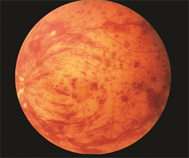
·      + papilledema/ swelling of the optic disc on fundoscopy
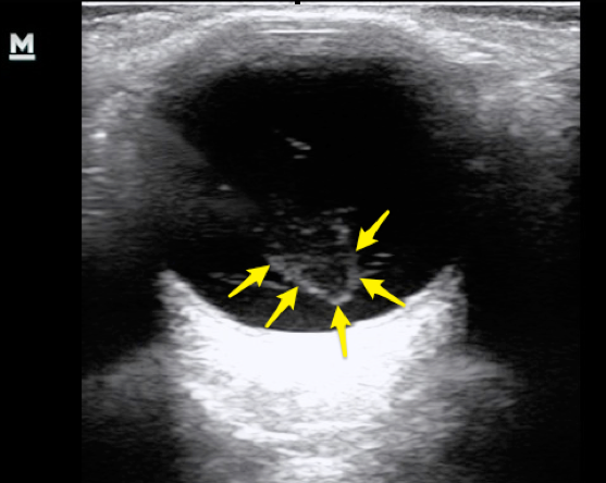
potd eye papill.jpg

·      optic sonography

potd us eye.jpg
  • ONSDs should be measured 3 mm behind the papilla, an average of less than 5 mm is considered normal.

  • ONSD > 5 mm has been shown to be 90% sensitive and 85% specific for ICP > 20.

·      Classic presentation: young, obese female

·      + association has been found with this diagnosis and the use of oral contraceptive medications, tetracycline, anabolic steroids, and vitamin A

·      Pathophysiology is not well understood but thought to be caused by an imbalance in CSF production and reabsorption

·      Diagnostic criteria include an alert patient with either a normal neurologic examination or findings consistent with papilledema, visual field defect, or an enlarged blind spot

·      Definitive dx: Lumbar puncture

  • done in a recumbent position reveals an elevated CSF opening pressure of more than 20 mm Hg in an obese patient (normal being up less than 20 mm Hg).

  • normal CSF analysis.

·      CT head may show “slit like” or normal ventricles without mass effect

·      DDx: glaucoma, venous sinus thrombosis, ICH, IC mass.

·      Treatment

  • Repeat LPs  

  • Acetazolamide

  • Surgical shunt if severe and refractory

  • offending agents such as oral contraceptive medications should be discontinued.

·      Permanent loss of vision can occur in up to 10% of patients, and higher if left untreated


Sources:


  • Dubourg J, Javouhey E, Geeraerts T, Messerer M, Kassai B. Ultrasonography of optic nerve sheath diameter for detection of raised intracranial pressure: a systematic review and meta-analysis. Intensive Care Med. 2011;37(7):1059-68. [pubmed]

  • Blaivas M, Theodoro D, Sierzenski PR. Elevated intracranial pressure detected by bedside emergency ultrasonography of the optic nerve sheath. Acad Emerg Med. 2003;10(4):376-81. [PDF]

  • https://www.ultrasoundoftheweek.com/uotw-5-answer/

  • Peer IX

 · 

POTD: Trauma Tuesday: Lateral Canthotomy

When to perform it?

·      To release orbital compression syndrome, most commonly due to retrobulbar hematoma.

·      IOP > 40, the pressure that indicated that you need to cut and release the compartment syndrome.

·      Without decompression, irreversible vision loss due to increasing orbital pressure may occur in as little as 90-120 minutes.

Clinical situation: trauma to the head/face.

Physical exam:

https://www.emra.org/emresident/article/emergency-department-evaluation-of-blunt-orbital-trauma/

https://www.emra.org/emresident/article/emergency-department-evaluation-of-blunt-orbital-trauma/

CT head and face

112-208-f2.jpg

familiarize yourself with the anatomy

potd anatomy eye.png

Before starting, highly consider sedation.

Steps:

1)    Generously inject 1% Lidocaine with epi to numb that lateral canthus to the orbital rim.

  • Do this by directing the needle away from the globe itself

  • Helps with bleeding and with pain.

2)    Using the needle driver as your hemostat, advance from the lateral canthus to outer orbit rim. Clamp down and hold for 1- 2 minutes.

3)    Using your small scissors, cut the lateral canthus to the orbital rim.

4)    Then cut inferiorly to cut the inferior crus of the lateral canthus (you may need to probe around to feel the structures)

5)    Repeat IOP. If the IOP is not immediately lower, then cut the superior crus of the lateral canthus and recheck pressure.

Pearls:

·      There is a lot of swelling. It can be hard to fit your hemostat in place and to feel your landmarks. Use you instruments to feel/probe around.

·      Also, do not worry about cutting too much. You are doing this to save this patient’s vision. After discussing this with optho, these are repaired quite easily days/weeks later. For example, Dr. Tome Levy performed this once on a patient that I later followed up with in split flow about 2 weeks later. There was no physical evidence on physical exam that this had ever happened. At first I thought I had the wrong patient in front of me… but the patient confirmed that a week and half ago he had in fact had the optho plastic surgery to repair it.  

Sources:

https://emedicine.medscape.com/article/82812-overview

http://www.tamingthesru.com/blog/annals-of-b-pod/ocular-emergency

This is an excellent emrap video that reviews the procedure: https://www.youtube.com/watch?v=tgQaKVGynFA

 · 

Do you see what eye see?

Do you see what eye see?

Eye complaints can be abundant in the ED - so what should we focus on? Today's pearl is going to focus on the diagnoses of atraumatic vision loss.

Let's break it down by symptoms: Painless or painful?

Good physical exam must include: visual acuity, visual fields, pupillary exam, fluorescein woods lamp, slit lamp and of course - a head to toe complete physical exam. 


Painless Vision Loss: 

  1. TIA/stroke: sudden vision loss usually from embolic or thrombotic event, can often be from carotid artery occlusion. If transient, often called amaurosis fugax but that can apply to any cause of transient vision loss. Risk factors: it is a stroke/TIA of the eye, so same cardiovascular risk factors. Testing: MRI, ECG, Echo, Carotid US. ED role: Don't miss it - this is a stroke and should admitted to a monitored stroke unit.

2. Central retinal artery occlusion: Sudden, permanent typically unilateral vision loss (permanent damage ~90 minutes of occlusion). Risk factors: atherosclerosisdisease (HTN, DM, Smoking, etc.) and treatment involves treating risk factors (varied success with laser treatment of pressure decreasing eye drops); or secondary to Giant cell arteritis (+/- headache, pain with hair burshing or chewing)  in which case rapid high dose corticosteroids can prevent vision loss. PE: decreased visual acuity, asymmetric red reflex, "Cherry red spot" on the macula on fundoscopy. ED role: urgent/emergent optho referral or ED consult. Recognize if cause is giant cell arteritis (CRP, ESR) and treat with high dose steroids. Consider timolol 0.5% topical drops but no strong evidence. Will need PCP to address concurrent risk factors. 

3. Central retinal vein occlusion: often times sudden but occasional gradual (days-weeks) vision loss due to swelling of the macula. Risk factors: same as above and glaucoma. Think of this as a DVT for the eye. PE: decreased visual acuity. "Blood and thunder" on fundoscopy. ED role: Evaluate for cause or presence of other pathologies secondary to a hypercoagulable state (DVT, PE, sickle cell, etc.). Refer to optho and will need PCP to address concurrent risk factors. 

retinal vein occlusion image-full.png

4. Retinal Detachment: Sudden, spontaneous "flasher and floaters" or "spots or stars" or loss of vision that is like a "curtain closing." Usually loss of peripheral vision. Risk factors: recent eye injury or surgery, severe nearsightedness (think elderly). ED Role: POCUS - look for a delicate floating line in the anechoic eyeball space. If their is no macular separation, their vision can be saved! This is an optho emergency. Unless you're Errel, err on the side of caution and call optho for all of these since we might not be able to definitively determine if the macula is involved.

5. Vitreous Hemorrhage and Posterior Vitreous Detachment: Floaters, strings, or cobwebs in their vision that change with eye movement. Flashes of light. Risk factors: retinal damage (surgery, trauma, prior retinal tear), Diabetic Retinopathy, trauma, sickle. ED role: POCUS: swirling cloud-like opacity at moves with ocular movement and is not tethered to the optic disk or retina "washing machine sign". ED role: Avoid Anticoagulation, elevate head of bed, optho referral.

6. Optic Neuritis/papillitis: painless vision loss over hours to days, typically unilateral. +/- prior episodes. +/- other neuro symptoms. May worsen with eye movement. Risk factors: typically females 18-45, multiple sclerosis. Viral: mono, zoster, encephalitis, TB. Physical exam: decreased acuity, relative afferent pupil defect, often normal fundoscopy; requires complete neuro exam. ED role: MRI brain. Diagnose underlying cause. Often required admission for continued IV high dose steroids. 

Painful Vision Loss:

1. Acute angle glaucoma: sudden, usually form bright to dark environment (movie theater). Often with headache, N/V, and light sensitivity/halos. PE: midsize, nonreactive pupil. Must include tonometry, IOP>20! Risk factors: Asian, femaile, shallow anterior chamber. ED role: timolol  0.05% 1-2 drops (beta blocker, watch for systemic absorption SE) eye drops and brimonidine (alpha agonist) eye drops to reduce IOP. Miotic agent pilocarpine (2-4% 1-2 drops q15 m). Titrate until symptoms improve, IOP decreases. Systemic carbonic anhydrase inhibitors like acetazolamide 500 mg IV. Elevate head of bed. Treat nausea and headache, too! Urgent optho referral.

2. Corneal Ulcer: foreign body sensation, red eye. PE: injected conjunctiva, gray patch on cornea. Increased fluorescein uptake on Woods lamp. Possible hypopyon purulent collection. ED role: Evaluate for foreign body. Consider HSV keratitis. Start opthalmic antibiotic drops, must have pseudomonas coverage for contact lens wearers (tobramycin), consider antifungal. **Admit for IV ceftriaxone for suspected gonococcal infection. Will need optho evaluation and ulcer culture. 

cornealulcer.jpg
Corneal-Abrasion.jpg

3. Uveitis/Iritis: painful, progressing red eye worse with eye movement, +photophobia suggests anterior. Floaters suggests posterior, often no pain. Can be panuveitis. Causes can be inflammatory (HLA B-27 usually bilateral, eg. reactive arthritic, psoriasis, IBD, ankylosing spondylitis; sarcoid, Kawasaki), traumatic, infectious (toxoplasmosis, CMV, HSV, adeno, measles, mumps, TB, syphilis, Lyme), or secondary to medications(sulfa). PE: injected conjunctiva with ciliary flush (erythema around the iris). Sluggish, constricted, or irregular pupils. Slit lamp shows cloudy anterior chamber and "cell and flare." ED role: evaluate for underlying cause (STD screen, CXR for TB, etc.) and refer to optho urgently and appropriate f/u. 

I am certain that this is not a comprehensive list, so please - learn on!

Buzzword pearls to part with: 

OpthalmologySession_web.jpg
 ·