Knee Dislocation

If you remember one thing from this post remember this: up to 50% of dislocations spontaneously reduce before presentation to the the ER - these patients are STILL at high risk for neuromuscular injury. Take a good history about the mechanism of injury, get a good exam, and follow the instructions below, to make sure you dont miss a popliteal artery or peroneal nerve injury!


Diagnosis of Knee dislocation:

  • up to 50% of dislocations spontaneously reduce before presentation to the ER, but that doesn’t mean a neurovascular injury didn’t occur during the dislocation

    • consider the mechanism of injury: motor vehicle accidents, other high velocity mechanisms (falls, downhill skiing, football) make dislocation more likely

    • in rare cases, low velocity injuries in the obese, or sudden twisting motions in athletes can also result in dislocation

  • knee exam should focus on appearance, integrity/stability of joint, distal perfusion, and evaluating for peroneal nerve injury

    • peroneal nerve provides ankle dorsiflexion, toe extension, and sensation to first dorsal web space

    • usually 3 or more major knee ligaments must rupture for the knee to dislocation, so any knee exam w/multi-planar instability should be a suspected dislocation that spontaneously reduced

  • Anterior dislocation is most common (50-60%) named for the direction of translation of the proximal tibia

  • Posterior dislocation is even more commonly associated w/popliteal artery injury

Picture1.png


Diagnosis of complications, especially in a spontaneously reduced knee:

  • most common injury is popliteal artery injury

    • presence of pulses does not exclude injury to the popliteal artery

    • if missed, can end up with AKA (delay of pop artery repair beyond 8 hrs invariably leads to limb amputation)

    • this is a highly litigated injury

  • ruling a popliteal injury via “hard signs” requires an immediate vascular surgery consult

    • hard signs include absence of pulse, pale or dusky leg, parasthesias and paralysis, rapidly expanding hematoma, pulsatile bleeding, bruit or thrill over the wound

  • There is no physical exam sensitive enough to rule out popliteal injury!!

    • quality of evidence behind ABI is poor as well

    • Wills et al prospective study suggests that normal ABI + period of observation w/no change in exam is 100% sensitive combination for ruling out vascular injury

    • Standard angiography is the standard of care

      • CT angio with runoff is next best test in the ER - ORDER THIS if concern for vascular injury


Management of a currently dislocated knee:

1. First and foremost the immediate reduction, and if neurovascular compromise exists – without radiographs.

a. Look for an anteriomedial skin furrow or “pucker sign” when the knee is extended – this signifies a posteriolateral dislocation, which are not reducible by closed reduction (require open reduction in OR).

Picture2.png

a. Document neurovascular exam before and after reduction attempts

b. Initial approach should be application of longitudinal traction to lower leg (anterior dislocation may require additional lifting of distal femure, while posterior may require lifting the proximal tibia to complete reduction.

Picture4.png


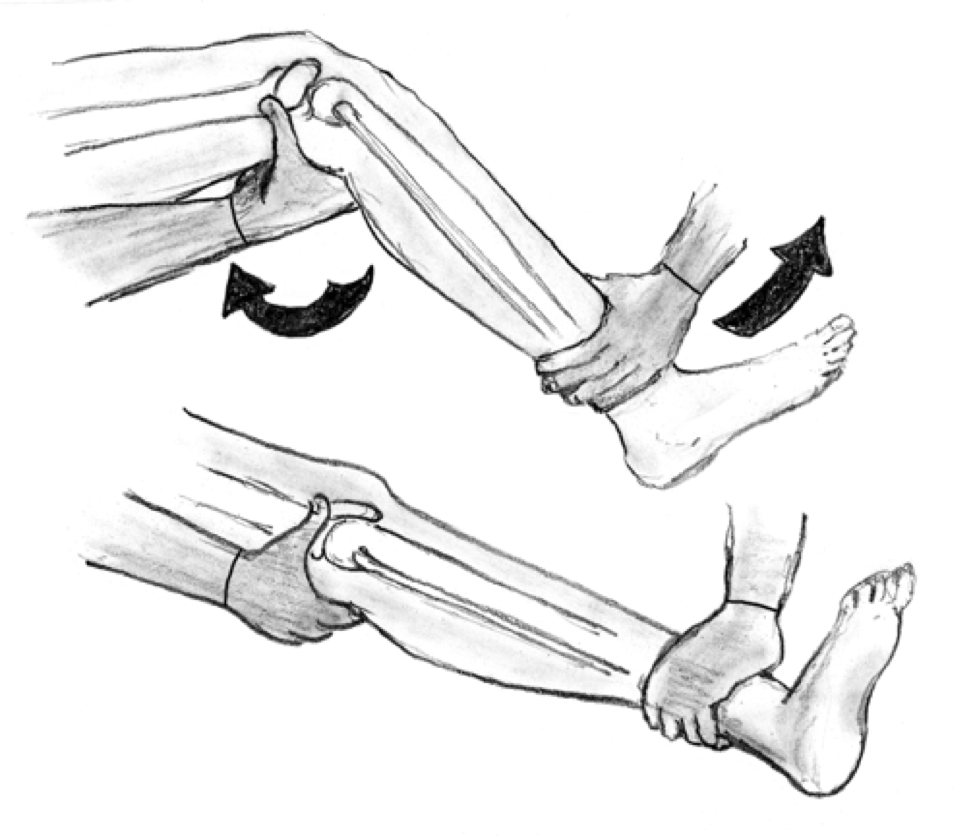
c. After reduction, the knee should be immobilized in a long leg posterior splint with the knee in 15-20 degrees of flexion

d. Again, these pts need either normal ABI with close monitoring and serial exams OR CTA to rule out vascular injury after initial reduction

References:

http://www.emdocs.net/knee-dislocation-pearls-and-pitfalls/

https://www.emrap.org/episode/emrap20203/medicallegal

 · 

Metacarpal Fractures

Trauma Tuesday!Metacarpal Fractures

mcp fractures.png


Why do we care so much about a few small bones in the hand? Because missed injuries can lead to permanent disabilities--we (as well as our patients) need our hands for pretty much everything.

How to assess for these injuries? Do your typical hand exam but pay special attention to:

Rotational alignment! Have the patient flex at the MCP and PIP, forming a loose fist with the DIPs extended (as in the figure below, to the left). The axis of each digit should merge near mid wrist. Rotational malalignment will cause deviation of this axis for the injured digit.

mcp fractures 2.png


Rotational malalignment is usually an indication for operative repair, so be sure to check for it. 

Don't forget: Any open wound over the MCP should alert you to the possibility of a "fight bite"--usually require exploration or washout. This needs EMERGENT ortho evaluation. 

Diagnosis

Get X-rays - AP, lateral, and oblique views; pay special attention to the lateral as this is what you will use to measure angulation.

For the quick and dirty: acceptable shaft angulation is 40° for 5th MC, 30° for 4th MC, 20° for 3rd MCP, and 10° for 2nd. Reduce if there is greater angulation. 

Management

NONOPERATIVE: For stable fractures, those without rotational deformities, and those with acceptable angulation and shortening (usually 2-5mm for each shaft) => nonoperative repair: 

Reduce a dorsally angulated neck fracture before splinting, usually done via the Jahss technique. (https://youtu.be/40irKoUJqsM)

For MCP head/neck/shaft fractures, radial or ulnar gutter splint depending on which MCP is injured. For MCP base fractures, wrist splint. 


OPERATIVE: For open fractures, intra-articular fractures, fractures with rotational malalignment, significantly displaced or angulated fractures, or in the event of multiple MCP fractures => operative repair

Err on the side of prompt orthopedic follow up. 


Sources

https://emergencymedicinecases.com/episode-29-hand-emergencies/

https://coreem.net/core/metacarpal-fractures/

https://www.orthobullets.com/hand/6037/metacarpal-fractures

 · 

Joint Aspiration: Ankle

When to tap?

When you have a debilitating ankle injury with swelling at the tibiotalar joint preventing range of motion at that joint ie: dorsiflexion/plantarflexion.

What about the differential?

Ankle arthrocentesis allows for rapid identification of septic arthritis vs. gout vs. pseudogout vs. osteoarthritis vs. rheumatoid arthritis.

What are your landmarks?

The goal is to avoid the Dorsalis pedal artery, the peroneal nerve and the tendon of the Extensor Hallucis Longus (EHL). It is recommended to use an anterolateral approach where the joint line can be found between the lateral edge of the EDL and the medial edge of the lateral malleolus (Yellow Arrow Image 1). Plantarflex the ankle while the patient is bent at the knee in the supine position to widen the joint space prior to performing the procedure.

IMAGE 1:

Foot.jpg


How do you perform it?

  • 1. Patient should be in a supine position with the ankle in plantar flexion with plantar surface flat on the bed.  

  • 2. Mark you landmarks (see above).

  • 3. Prepare the site (ex. chloraprep)

  • 4. Anesthetize the area with smaller needle(23/25 gauge) creating a wheal and then advance creating the start of a projected path towards the joint capsule.

  • 5. Attach a 5 or 10 cc syringe to a 20 or 22 gauge needle and advance the needle into the joint space pulling negative pressure as you advance. The needle should be directed perpendicular to the tibia. If your syringe starts to fill up, and you need to get more fluid out, change out your syringe using hemostats to hold the needle. Most wrist and ankle effusions will yield only 1-3cc of fluid.

What about Ultrasound Guidance?

YES. This can absolutely be used to assist you in performing the procedure and will allow for visualization of your needle tip during aspiration.


For ultrasound guidance an anteromedial approach is generally used.

Landmarks- Place probe in between the TA tendon and EHL tendon, then rotate longitudinally with the probe marker facing the patient’s head  (Blue Arrow IMAGE 1). You will actually be inserting your needle medial to the TA tendon (Red Arrow IMAGE 1).

Image 2: 

more-tips-and-tricks-6.jpg
more-tips-and-tricks-7.jpg

Image 3:

AnkleTap.png