Acute Compartment Syndrome

Acute compartment syndrome is when the pressure in a muscle compartment increases, compromising circulation and function. This occurs because the compartment is surrounded by a fascial membrane that restricts further expansion. It typically occurs after trauma, crush injury, or burns. Signs include severe pain (earliest sign), pallor, paresthesia, paresis, and pulse deficit. To measure compartments, you take your measurement device and insert it into the compartment of interest. 

How to set up your measurement device: 

  • Your materials include a sterile 3cc saline syringe, chamber, and needle. The needle has a side port (hole) for measuring pressure.

  • Connect the syringe, chamber, and needle

  • Flush the chamber and needle with saline to get rid of the air; do this by holding the entire device at a 45-degree angle.

  • Load into the monitoring unit and press zero, you should see 00

  • Insert the needle into the compartment of choice and hold it for reading

There are two ways to assess for compartment syndrome. You can use the absolute or delta pressure (normal: 0-8). Suspect compartment syndrome if:

  • the absolute pressure is > 30 mmHg

                                   OR

  • The delta pressure is < 30 mmHg

    • Delta pressure = diastolic pressure - compartment pressure. This means that the pressure in your compartment is so high that it is close to your diastolic blood pressure

Tip: remember the number 30 

These patients require a fasciotomy so call ortho ASAP. Meanwhile, you should level their affected limb and support BP if hypotensive to help maintain perfusion. 

Thanks for reading!

-Ariella

References: 

https://www.emrap.org/episode/trauma1/compartment

https://www.emrap.org/episode/measuring/measuring


POTD: Umbilical Vein Catheterization

I wanted to review a fairly rare but lifesaving EM procedure in neonates. This procedure is done fairly commonly in the NICU/L&D, but is done less frequently in the ED, especially with our excellent nurses who can literally get the most impossible venous accesses. If you went to Airway day, you might recall Dr. Sokolovsky describing her harrowing tale of providing neonatal resuscitation at Burning Man and performing an umbilical vein catheterization with an 18-gauge IV. Super wild! So for anyone who might find themselves in a similar poop-inducing situation with no pediatric support or NICU available, this is for you!


Umbilical vein catheterization is indicated in a neonate within 14 days post-birth requiring IV resuscitation. The stump must be "fresh", so it is most ideal in the newly born neonate. Here is an excellent video overviewing the following steps. https://pedemmorsels.com/wp-content/uploads/2019/08/UVC....mp4

Here's what you'll need:

  • Sterile gloves (gown and drape less non urgent)

  • chlorhexidine

  • forceps

  • scalpel

  • umbilical line (5 French is standard, 3.5 French in very premature baby)

  • three-way stopcock

  • umbilical tape of 3-0 silk/nylon

  • NS flush

In peds, we have umbilical vein catheterization trays located on the top shelf in Bay 31 that includes all of the above except the catheter. While the umbilical line is the traditional teaching, you can use any tube that can fit into the vein - that means an 18 gauge IV, pediatric central line, feeding tube, etc


Prep the umbilical stump

  1. Flush the line and place

  2. Sterilize the entire umbilical stump, including the clamp at the end of the stump, and the abdomen

  3. Tie the umbilical tape (or a silk string) around the base of the stump loosely. This helps decreased blood flow for when the clamp is eventually removed. It can also be tightened to secure the line once placed

  4. Holding the clamp, make a transverse cut off the stump to remove the distal tip. Cut should be made directly below the clamp or 2 cm from the abdomen.

Identifying umbilical vein and prep for insertion

  1. Identify the umbilical vein. The anatomy of the stump involves two smaller umbilical arteries and one umbilical vein. The arteries are typically smaller and thicker lumen, while the vein is larger and more collapsible (see below)

  2. Remove any clots from the vein and gently dilate the vein with forceps

  3. gently insert the line, when you get blood return insert 1-2 cm deeper, or approx 3-5 cm. If there is resistance, consider loosing the umbilical string.

  4. Aspirate blood and flush with NS. Secure the line by tightening the umbilical string and securing with tape or purse string suture

Complications of UVC placement are similar to CVP placement: excessive bleeding, infection, thrombosis, arterial insertion. Specifically to UVC is risk of insertion too deep into the portal venous system or right atrium, which can lead to hepatic necrosis and perforation.

Resources:

https://first10em.com/umbilical-vein-catheterization/

https://wikem.org/wiki/Umbilical_vein_catheterization

https://www.ncbi.nlm.nih.gov/books/NBK549869/


POTD: Primary Hypothermia!

With the turn of the weather and puffy jacket season in full throttle, let's review hypothermia. In this POTD, we're going to be focusing on primary (environmental) hypothermia, and not hypothermia due to secondary causes such as sepsis, hypothyroidism, metabolic derangements, or trauma. 


Hypothermia is defined as core temperature < 35C


  • between 1991-2011, an average of 1,300 deaths/year in the US attributed to environmental hypothermia

  • At risk populations: elderly, lack of shelter, alcohol/drug abusers, frequent exposure (winter sports)


The modified staging system for hypothermia classifies severity into 4 categories, of which the chart below summarizes and demonstrates the general clinical picture


Initial evaluation should include the following:

  • Basic vital signs including core temperature

  • BGM (hypoglycemia)

  • EKG (bradycardia, Osborne waves, arrhythmias)

  • Basic labs (hypokalemia)

  • lactate, CK (rhabdomyolysis)

  • coags and fibrinogen (DIC)

  • consider TSH, cortisol, 


Management of ABCs:

Airway/Breathing

  • Intubate as necessary, however encourage holding off intubation as patients are often hypotensive and prone to arrhythmias. Attempt rewarming and stabilization prior to hemodynamic stressors of intubation.

Circulation

  • bradycardia 

    • hypothermia itself causes bradycardia, thus the key to treating bradycardia is rewarming. Giving medications to speed up HR is generally not encouraged and can induced arrhythmias

  • hypotension 

    • again, often hypothermia induced, may also be secondary to bradycardia. Rewarming should improve pressures, and vasopressors generally discouraged as it can also induced arrhythmias

    • BUT - vasodilation can occur with rewarming, so if there is a drop in pressure, vasopressors should be considered

  • access 

    • if central access is needed, keep the guidewire shallow to prevent entry into the right ventricle. The hypothermic heart is incredibly sensitive to VT/VF, so don't go tickling that ventricle. Femoral access or midline access is preferred
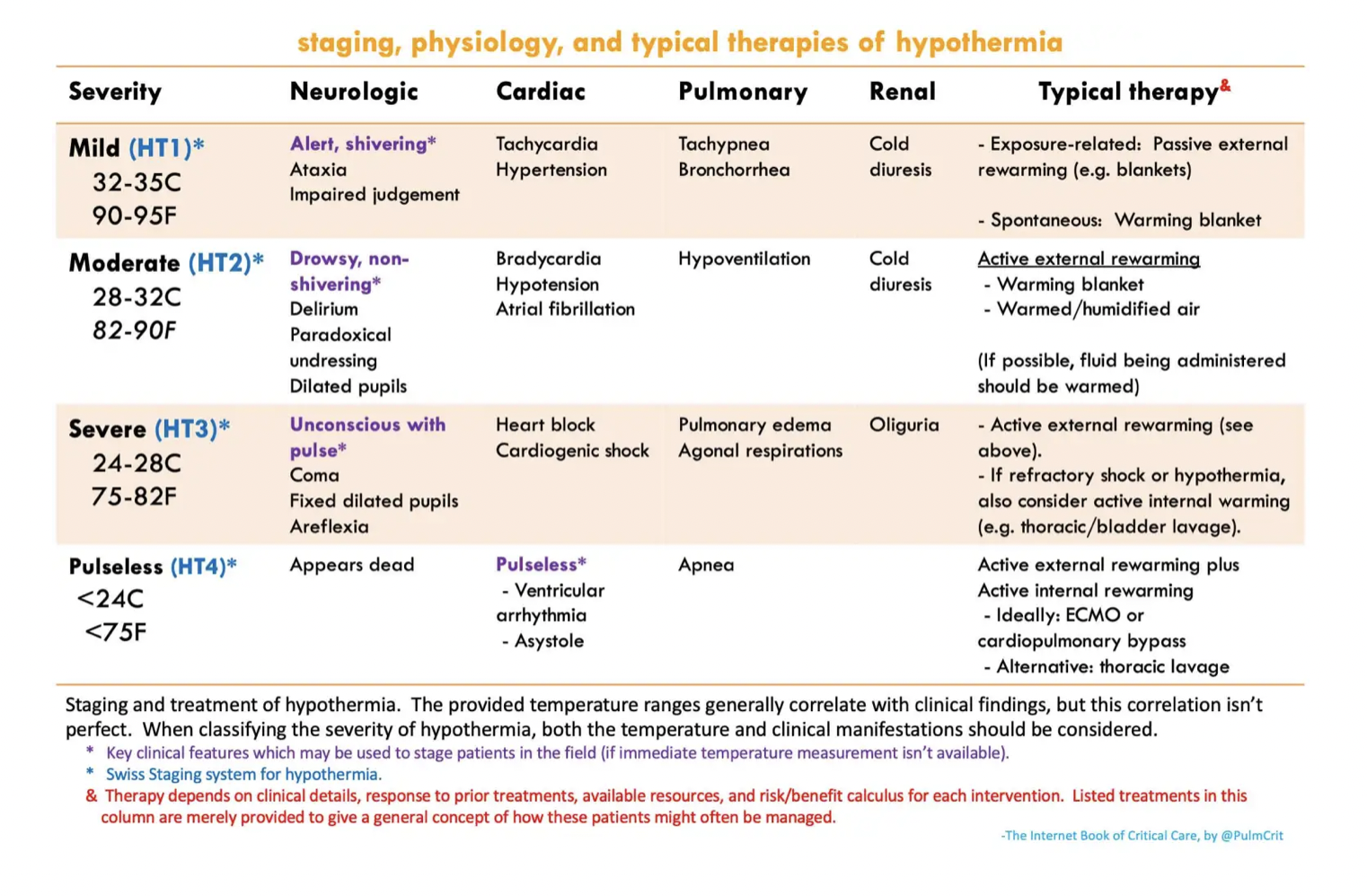

EKG in hypothermia:

Patient may initially present with tachycardia, however as hypothermia sets in, everything gets slower. Just imagine taking a NSR rhythm stripe and click-dragging it horizontally. You often see bradycardia, prolonged QTC, prolonged PR, and the presence of Osborne waves. 

  • Osborne waves (also known as J waves) - a deflection at the junction of the QRS and ST segments, present usually temp < 30C. The size increases with worsening hypothermia

The most common dysrhythmia is atrial fibrillation, but cardiac arrest is due to eventual VF/VT or asystole

Management of hypothermia in the pulseless patient:

The famous phrase: "They aren't dead unless they're warm and dead" indeed is true. To be considered dead, the patient's core temperature should be ~32C before calling it.

ACLS: Modifications in setting of hypothermia

  • Delayed or intermittent CPR can be adequate due to low oxygen demand. Some say for T < 28, there can be pauses of <5 minutes CPR after every 5 minute interval of CPR if necessary. 

  • Medications may fail to metabolize and will accumulate in the system, therefore prolonged intervals between epinephrine pushes and limiting repeated doses are recommended. 

  • Similarly, defibrillation is poorly effective in T < 30C and repeated defibrillation may induce myocardial damage. 

    • Suggested treatment by AHA: Can attempt one defibrillation if VF/VT present, otherwise hold further defibrillations and all IV medications until core T > 30C

Internal Rewarming:

  • ECMO: The best option for sever hypothermia. It allows for better organ perfusion, active rewarming as much as 7-10C per hour, and allows you to stop compressions. As an ECMO center, we should definitely consider getting ECMO downstairs to cannulate.

  • Thoracic Lavage: achieved through placement of two chest tubes, left preferred over right if only able to place one tube. Can achieve 3-6C warming/hour. Use Belmont or warm tap water if available, and monitor appropriate ins and outs to prevent tension PTX from improperly draining tubes

  • Bladder Lavage: less effective, however is easier to implement. Use dedicated 3-way Foley catheter or consider instilling 300 cc warmed fluids, hold for 15 minutes, and draining bladder. Rinse and repeat.

Management of hypothermia in patient with pulse:


  • External rewarming: Removing cold/frozen clothes and fully wipe down of snow. Employ external rewarming with Arctic Sun (preferred due to direct contact with skin to improve rewarming), warm blankets or Bair Hugger. 

  • Respiratory rewarming: Warmth is typically lost through expirations. Therefore consider rewarming via respiratory support. In non-intubated patients, HFNC or CPAP/BIPAP with highest temperature settings. Intubated patients should have heated and humidified air set up with the ventilator (ask respiratory)

  • Fluid rewarming: hypothermia induced "cold diuresis," therefore patients often require volume resuscitation. Warm IV fluids, ideally crystalloid, should be administered. Keep in mind that this method prevents further dropping temperatures, but is ineffective for raising core temperature. 

  • Lavage can also be considered in moderate/severe hypothermia in patients with pulses - see above.


Resources:

https://emcrit.org/ibcc/hypothermia/

http://www.emdocs.net/em3am-hypothermia-2/

https://www.saem.org/about-saem/academies-interest-groups-affiliates2/cdem/for-students/online-education/m4-curriculum/group-m4-environmental/hypothermia

https://wikem.org/wiki/Accidental_hypothermia