POTD: Subcutaneous insulin in DKA management

Happy to start off this month of Admin Resident with a requested POTD, the SQuID trial (Subcutaneous Insulin in Diabetic Ketoacidosis).


Diabetic Ketoacidosis (DKA) management traditionally involves intensive treatments like fluid resuscitation, electrolyte replacement, and intravenous insulin infusions. This approach often necessitates ICU admissions, especially for mild to moderate DKA cases, due to hospital policies. However, the strain on ICU resources and the extended ED stays prompt the need for alternative treatments. A promising solution emerges with the use of Subcutaneous (SQ) insulin in mild to moderate DKA, potentially reducing ICU admissions and ED overcrowding.


Clinical Question:

Does the SQ insulin protocol reduce ED length of stay in adult patients with mild to moderate DKA compared to traditional IV infusion?

Methodology:

    • Approach: Implementation of the SQuID (Subcutaneous Insulin in Diabetic Ketoacidosis) protocol in an urban academic ED.

    • Study Design: Prospectively derived, quasi-experimental (pre-post) study, with retrospective data analysis.

    • Participants: Adult ED patients with mild to moderate DKA.

    • Exclusions: Severe DKA (HCO3 <10mmol/L, Arterial pH < 7.0), pregnancy, serious infections, and other critical conditions.

Outcomes Measured:

    • Fidelity: Frequency of required glucose checks every 2 hours.

    • Safety: Proportion of patients needing rescue dextrose for hypoglycemia.

    • Operational Impact: ED Length of Stay (LOS) and ICU Admission Rate.


Results:

    • ED LOS Reduction: The SQuID protocol showed a reduction in ED LOS compared to the traditional method.

    • ICU Admissions: Slight decrease, though not statistically significant.

    • Safety: Comparable between SQuID and traditional protocols.


Utility in the Emergency Department

The SQuID protocol's primary advantage in the ED is its potential to significantly reduce patient LOS. This efficiency can alleviate the overcrowding issues, allowing the ED to manage other emergent conditions more effectively. However, successful implementation requires careful planning and education, as it alters the conventional treatment approach for DKA.


Pitfalls to Consider

While promising, the SQuID protocol has several pitfalls:

    1. Increased Monitoring Requirement: The protocol demands more frequent glucose monitoring, which could strain ED resources.

    2. Misclassification Risk: Incorrect assessment of DKA severity could lead to inappropriate protocol application.

    3. Education and Training Needs: Extensive education for ED staff, hospitalists, and nurses is necessary for effective and safe implementation.

    4. Safety Concerns: The increased incidence of hypoglycemia events, compared to historical controls, necessitates vigilant monitoring.


Strengths and Limitations:

    • Strengths: Addresses a critical clinical question with a clearly defined protocol.

    • Limitations: Includes potential for increased glucose monitoring frequency, subjective decision-making affecting LOS and ICU admissions, and limited generalizability due to the single-center setup.


Discussion: Implications and Considerations

The SQuID protocol demonstrates a potential shift in DKA management. It not only reduced ED LOS but also showed equivalent safety compared to the traditional insulin infusion pathway. However, several challenges like the need for extensive hospital-wide education, potential misclassification of DKA severity, and concerns about hypoglycemia management necessitate cautious implementation.

Clinical Take Home Point

The SQuID protocol offers a promising alternative for managing mild to moderate DKA, potentially reducing ED LOS and ICU admissions. However, considerations regarding hospital-wide education, monitoring frequency, and safety concerns mean that it's not yet ready for widespread adoption.

References

    • Griffey RT et al. "The SQuID Protocol (Subcutaneous Insulin in Diabetic Ketoacidosis): Impacts on ED Operational Metrics." Acad Emerg Med 2023. PMID: 36775281

 · 

POTD: Medical Trivia Potpourri

 ·   · 

Why is baby aspirin 81mg instead of 80mg?

This is a remnant of the medieval apothecary system. In the late 1700s, medication dosages in the apothecary system were based on the weight of a grain of barleycorn, with the unit of weight termed the grain (abbreviated gr)1. The standard dose of aspirin used back then was 5 gr, equivalent to 325mg. This is also why Tylenol tablets also comes in multiples of 325mg. Baby aspirin was one quarter of that, which is equivalent to 81mg.

Where does the term “mad as a hatter” come from?

Poisoning. This comes from mercury poisoning. In the 18th and 19th centuries, mercury nitrate was used in the process of turning animal fur into felt products. Many hatters developed tremors and neuropsychiatric symptoms during this period2.

How glucagon received its name.

Kimball and Murlin found that a substance secreted from the pancreas causes hyperglycemia when injected into rabbits and dogs. They decided to name this substance glucagon, short for “glucose agonist3.”

What animal venom contributed to research in GLPs?

Gila monster. In the 1990s, Dr. Eng was researching in how Gila monsters were able to maintain blood sugar despite long periods of not eating, and discovered a peptide called exendin-4 in its venom, this is structurally and functionally similar to GLP14.

Adrenaline vs epinephrine? Acetaminophen vs APAP?

It’s because the English language is derived from so many different roots. Adrenaline derives from Latin, “ad + renal”, or “on kidney”. Epinephrine derives from Greek, “epi + nephros”, which again translates to “on kidney.” The names from Tylenol comes from different abbreviations of its chemical structure.

  • Acetaminophen = N-acetyl aminophenol.

  • Paracetamol = N-acetyl-para-acetyl-amino-phenol.

  • APAP = N-Acetyl-Para-Acetyl-Amino-Phenol.

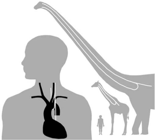
You think you’ve got a lot of (laryngeal) nerve?

The recurrent laryngeal nerve branches from the vagus nerve (CN X) to innervate much of the larynx and control speech. Due to embryological development, the left and right laryngeal nerves wrap around the aortic arch and right subclavian vessels, respectively. As we develop, the heart grows further away from our throat, causing stretching of the “recurrent” nerve. This leads the nerve to make a 10cm U-turn in humans. In Giraffes, the recurrent laryngeal nerve approaches 5 meters. In dinosaurs, this is hypothesized to reach 28 meters5.

Was “War-farin” discovered during research as part of some war effort?

Close, but not quite. Actually, it's not really close either. Warfarin was discovered because of cows randomly bleeding to death. Cattle farmers invited researchers from the University of Wisconsin to figure out why. These researchers discovered that the cattle were eating moldy sweet clover hay, which was found to contain a substance called “coumarin” that anticoagulated the cows and caused them to hemorrhage. The patent rights for this discovery were given to the Wisconsin Alumni Research Foundation (WARF) which is where “WARFarin” originates from, as well as the generic name “Coumadin” from the substance coumarin6,7.

References

1.           Zupko RE. Medieval Apothecary Weights and Measures: The Principal Units of England and France. Pharm Hist. 1990;32(2):57-62.

2.           Where did the phrase “mad as a hatter” come from? HISTORY. Published May 8, 2023. Accessed January 1, 2024. https://www.history.com/news/where-did-the-phrase-mad-as-a-hatter-come-from

3.           Müller TD, Finan B, Bloom SR, et al. Glucagon-like peptide 1 (GLP-1). Mol Metab. 2019;30:72-130. doi:10.1016/j.molmet.2019.09.010

4.           Exendin-4: From lizard to laboratory...and beyond. National Institute on Aging. Published July 11, 2012. Accessed January 1, 2024. https://www.nia.nih.gov/news/exendin-4-lizard-laboratory-and-beyond

5.           The “Unintelligent Design” of the Recurrent Laryngeal Nerve. Office for Science and Society. Accessed January 1, 2024. https://www.mcgill.ca/oss/article/student-contributors-did-you-know-general-science/unintelligent-design-recurrent-laryngeal-nerve

6.           Warfarin Discovery | Wisconsin Alumni Association. Accessed January 1, 2024. https://www.uwalumni.com/news/warfarin/

7.           Ankit’s brain. Pharmacy Selective Rotation.

 · 

Trialysis Length

During a recent shift, Dr. Waters asked me “can I use a the 15cm trialysis line in the femoral vein?” To which I replied “I don’t see why not.” That got me thinking, is there a reason why not? We do have two length catheters after all (15cm and 24cm). Low and behold there is a reason why not to use the shorter length trialysis catheter there. The reason is that the catheter will not make it past the lumen of the common iliac vein and into the IVC where it is recommended the end of the catheter sit. CVCs pose certain risks such as CRBSIs, DVTs, and vessel stenosis. There are even case reports of vessel erosion happening when a catheter sits in the iliac and not in the IVC.

Bottom line; use the right size kit for the appropriate vessel, like Dr. Waters eventually did! The rest of this post is an overview on trialysis catheter placement.

Trialysis catheter placement in the emergent setting is a procedure undertaken to quickly establish vascular access for hemodialysis in critically ill patients with acute kidney injury or end-stage renal disease. This intervention becomes necessary when traditional vascular access methods, such as peripheral intravenous catheters or arteriovenous fistulas, are not feasible or fail to provide adequate blood flow for dialysis.

Indications:

Urgent Hemodialysis: In cases of severe acute kidney injury or end-stage renal disease, where immediate initiation of hemodialysis is required. “AEIOU” acidosis, refractory hyperkalemia, ingestion of certain substances (methanol, ethylene glycol, lithium, salicylates), overload of volume refractory to medical management, and uremia.

Contraindications:

  1. Vascular Anomalies or Injuries: Presence of significant vascular anomalies or injuries at the potential catheter insertion site.

  2. Local Infections: Infection at the proposed catheter insertion site.

  3. Severe Coagulopathy: Placement may be contraindicated in patients with uncontrolled bleeding disorders. (Relative contraindication – should use more compressible sites like the femoral vein)

Equipment Needed:

  1. Trialysis Catheter Kit: Includes the catheter, guidewires, dilators, and sheaths.

  2. Ultrasound Machine: To assist in locating suitable veins and ensuring proper catheter placement.

  3. Sterile Drapes and Gloves: To maintain aseptic conditions during the procedure.

  4. Local Anesthetic Agents: For numbing the catheter insertion site.

  5. Syringes and Needles: For administration of local anesthetic agents and other medications as needed.

  6. Suture and Dressing Materials: For securing the catheter in place and maintaining a sterile environment post-placement.

Procedure:

  1. Patient Assessment: Evaluate the patient's clinical status, coagulation profile, and vascular anatomy to determine the most appropriate site for catheter placement.

  2. Informed Consent: Obtain informed consent from the patient or their legal representative, explaining the risks and benefits of the procedure.

  3. Preparation: Position the patient appropriately, and ensure sterile conditions using drapes and gloves.

  4. Local Anesthesia: Administer local anesthesia at the proposed catheter insertion site.

  5. Ultrasound Guidance: Use ultrasound to locate a suitable vein and guide the catheter insertion, ensuring proper placement.

  6. Needle placement: With a needle attached to a syringe, insert the needle and begin withdrawing on the syringe while progressing, both to see when blood returns, and to ensure no introduction of air bubbles. Needles should be at a 45 degree angle when inserted. Preferably there is ultrasound visualization of the needle inside the lumen of the vessel.

  7. Guidewire insertion: Remove the syringe from the needle and progress the guidewire. This should be able to occur smoothly. If it is not progressing smoothly, you may need to drop the angle of the needle, as the guidewire may be getting forced against the backwall of the vessel. The wire should only go in about 20cm.

  8. Confirmation of guidewire: Ultrasound visualization should be done to confirm the guidewire. Never assume the guidewire is in the right spot. Know it is, by seeing it is. This should be done in both short plane and longitudinal plane.

  9. Incision: Incise at the site of the guidewire to be able to dilate and place catheter.

  10. Dilate (twice): The trialysis catheter requires double dilation given how large the catheter is. Place the dilator over the guidewire (without letting go of the wire). Go approximately halfway down the dilator, remove the dilator, and then repeat with the next dilator. (Be mentally prepared for a fair amount of blood).

  11. Catheter Insertion: Introduce the catheter through the dilated tract, securing it in place using sutures.

  12. Confirmation: Confirm proper catheter placement using imaging techniques such as fluoroscopy or ultrasound.

  13. Post-Procedure Care: Apply a sterile dressing, monitor for any complications, and secure the catheter to prevent accidental dislodgement.

Sources:

  1. National Kidney Foundation. (2006). "Clinical Practice Guidelines for Vascular Access." Retrieved from https://www.kidney.org/sites/default/files/docs/12-50-0210_jag_dcp_guidelines-va_oct06_sectiona_ofc.pdf

  2. American Society of Nephrology. (2006). "Clinical Practice Guidelines for Hemodialysis Adequacy, Update 2006." Retrieved from https://www.kidney.org/sites/default/files/docs/12-50-0900_anemiaworkbook_upd-0926.pdf

 ·