Trauma Tuesday: Electrocution Injuries

 Epidemiology

-       3 primary age groups

o   Toddlers – household sockets, appliances, etc.

o   Adolescents – risk-taking behavior

o   Adults – occupational hazard

-       Lightning strikes – account for 50-300 deaths per year in US (mostly Florida)

-       ~6,500 injuries and 1,000 deaths annually from all electrocution injuries


Classification

-       Low voltage: ≤1000 volts (V)

o   Household outlets in US typically 120 V

-       High Voltage: >1000 V

o   Power lines > 7000 V

-       Alternating current (AC) = electrical source with changing direction of flow  household outlets

o   Induces rhythmic muscle contraction  tetany  prolonged electrocution as individual is locked in place

o   Although generally lower voltages, can be more dangerous than DC as the time of electrocution is much higher

-       Direct Current (DC) = electrical source with unchanging direction of current of flow  lightning strikes, cars, railroad tracks, batteries

o   Usually induces a single, forceful muscle contraction  can throw an individual with significant force  higher risk of severe blunt trauma 


Mechanisms of Injury

-       Induced muscle contraction  rhabdomyolysis

-       Blunt trauma

-       Burns

o   Internal thermal heating – most of damage caused by direct electrocution

o   Flash/Arc burns – electricity passes over skin causing external burns

o   Flame – electricity can ignite clothing

o   Lightning strikes can briefly raise the ambient temperature to temperatures greater than 54,000F


Severity of Injury – is determined by…

-       Type of current – AC vs. DC

-       Duration of contact

-       Voltage

-       Environmental circumstances (rain, etc.)


Clinical Manifestations

-       Cardiac – 15%, mostly benign and occur within few hours of hospital stay

o   Arrhythmias - Most occur shortly after the event, though non-life-threatening arrhythmias can occur a few hours after the event and are usually self-resolving. Generally, …

§  DC = asystole

§  AC = ventricular fibrillation

o   Other EKG findings – QT prolongation, ST elevations, bundle branch blocks, AV blocks, atrial fibrillation

-       Pulmonary

o   Respiratory paralysis – diaphragmatic muscle

o   Blunt trauma – pneumothorax, hemothorax, pulmonary contusions, etc.

-       Neurologic – generally, patient can APPEAR DEAD but is the cause of neurologic electrocution and may be temporary. IE.

o   Coma

o   Fixed, dilated pupils

o   Dysautonomia

o   Paralysis or anesthesia

-       Renal – Rhabdomyolysis

-       Skin – All kinds of burns

-       MSK – from severe muscle contractions

o   Always assume C-spine injury

o   Compartment syndrome

o   Fractures/Dislocations


Management – we’ll divide them into categories of severity. Basically, always do an EKG!!


1)    Mild (<1000V) – examples include brief house outlet shock, stun gun

a.     EKG – other work-up such as troponin and CPK usually unnecessary

b.     If history/physical unremarkable (patient endorses brief contact with house outlet) patient can be discharged without further work-up

c.     If PMH puts patient at higher risk of arrhythmia (cardiac disease, sympathomimetics) can do a brief period of telemetry observation

d.    Can always observe 4-8 hours to be on the safe side

e.     High Risk Features

                                               i.     Chest pain

                                             ii.     Syncope

                                            iii.     Prolonged exposure

                                            iv.     Wet skin

2)    Severe Electrocution (>1000V) – industrial accidents, lightning strikes

a.     Coding – pursue usual ACLS

                                               i.     Keep in mind traumatic causes of arrest (tension pneumothorax, etc.)

                                             ii.     KEY FACT: remember that patients with fixed, dilated pupils, no respiratory effort, and no spontaneous movement may only have TEMPORARY neurologic stunning

                                            iii.     Pursue resuscitation longer than usual as patient with ROSC can still have good outcomes  does not appear to be any definitive guidelines on when to terminate, at physician discretion

b.     Otherwise, broad medical and traumatic work-up and likely admission for telemetry monitoring (basically just send all the labs and images)

                                               i.     Start with primary/secondary trauma survey and further imaging as required

                                             ii.     Don’t forget CPK to assess for rhabdomyolysis

c.     Consider transfer to burn center


TL;DR

-       Treat as you would a trauma/burn patient

-       Most household outlet shocks – history/physical, EKG, and likely quick discharge unless high risk features

-       Industrial shocks – at best admit for telemetry. At worst prolonged ACLS as good outcomes are possible. Don’t forget traumatic causes such as tension pneumothorax


http://brownemblog.com/blog-1/2020/4/14/acute-care-of-the-electrocuted-patient

http://www.emdocs.net/electrical-injury/

http://www.emdocs.net/em3am-electrical-injuries/

http://www.emdocs.net/em-cases-electrical-injuries-the-tip-of-the-iceberg-view-larger-image/

https://www.tamingthesru.com/blog/air-care-series/electrocution


 · 

POTD: Tick Bites

 Tick Removal – there are multiple tips and tricks to do this, but most sources suggest…

-       Using a pair of tweezers (or forceps) and attempting to grasp the tick as close to the skin surface as possible

-       Pull upwards with gentle, steady traction. Do not jerk or twist

-       Do NOT squeeze, crush, or puncture the body of the tick – this may expel infectious contents

-       After removing the tick, wash skin thoroughly with soap and water


What to do if mouth parts remain in the skin?

-       UpToDate says to leave it in and they’ll be expelled on their own

-       WikEM says to excise under local anesthesia… seems aggressive


Important Ticks for Identification – The CDC has a good guide. If you’re squeamish with bugs, you’ve been warned and please skip this part. There are 3 main types of ticks found in the US.


1)    Ixodes Scapularis or “deer ticks” = LYME DISEASE. Other ticks do not transmit Lyme disease


-       Brown, about the size of a poppy seed but can be larger when engorged

-       Primarily found in the North-East and Midwest, less commonly in the Western US

-       Most famously transmits Lyme Disease, also anaplasmosis, babesiosis


2)    Dermacentor species or “dog ticks”

-       Brown with a white collar, about the size of a pencil eraser 

-       Primarily found in the Rocky Mountain States (Colorado, Idaho, Montana, Nevada, Utah, Wyoming, etc.)

-       Most known for transmitting, you guessed it, Rocky Mountain Spotted Fever

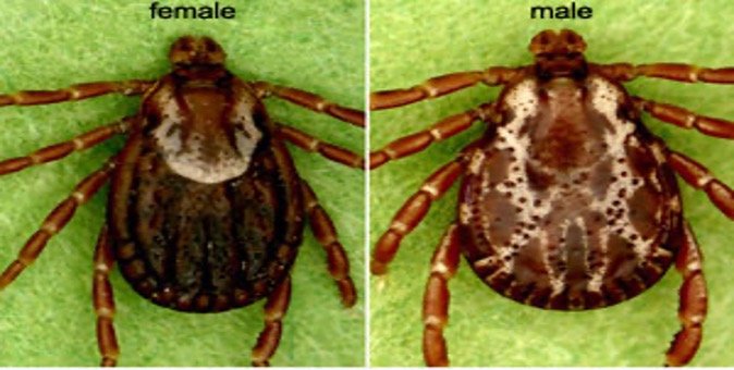
3)    Amblyomma Americanum or “Lone Star Tick”

-       Brown or black with a white splotch

-       Primarily found in the South, but can also be found in the Eastern US

-       Most known for Southern Tick-associated rash illness (STARI) and ehrlichiosis


Who needs prophylaxis? IDSA recommends prophylaxis only if ALL OF THESE CRITERIA ARE MET. It should be specified that this is for prophylaxis against Lyme Disease only.

-       The tick is identified as a deer tick

-       Tick is estimated to have been attached >36 hours or engorged (it takes time for the bacteria to exit the gut of the tick and enter the bloodstream). Ticks found crawling on skin automatically do not count.

-       The antibiotic can be given within 72 hours of tick removal

-       The bite occurs in a geographic location that Lyme Disease is highly endemic (can be found on CDC website)

-       There is no contraindication to take doxycycline (primarily appears to be hypersensitive or children < 8). If there is a contraindication, no second-line antibiotic exists


The prophylaxis is a single dose of 200mg doxycycline, or 4mg/kg up to a max of 200mg for children.

Antibiotic treatment following a tick bite is not recommended as a means to prevent anaplasmosis, babesiosis, ehrlichiosis, Rocky Mountain spotted fever, or other rickettsial diseases. Rather, patients should be warned and be vigilant against symptoms such as fever, rash, or other symptoms concerning for these diseases.

https://www.cdc.gov/ticks/tickbornediseases/tickID.html

https://www.uptodate.com/contents/what-to-do-after-a-tick-bite-to-prevent-lyme-disease-beyond-the-basics

https://wikem.org/wiki/Tick_borne_illnesses

https://wikem.org/wiki/Tick_removal


 · 

Mountain Sickness

Mountain sickness

Background: At higher altitudes, there is less oxygen. For example, at 10,000 feet, the air is 14% oxygen while at sea level in NYC, we are breathing in 21% oxygen. Mountain sickness is the manifestation of the body’s response to hypoxia. 

Clinical features

Usually only occurs in altitudes greater than 8000 ft unless patients are particularly susceptible to hypoxia (COPD, anemia). This is also why when flying, airplane cabins are usually pressurized to 7-8000 ft. Patients who have experienced altitude sickness are more likely to have repeat episodes when returned to the same altitude. A quicker rate of ascent is also more likely to lead to mountain sickness. Most often presents the 1st night or 2nd night at higher elevations. The average duration of symptoms in cases that self resolve is one day (the body successfully acclimates). 

Clinical criteria (most are CNS symptoms since the brain is most sensitive to hypoxia): An individual above 8000 feet presents with headache and one of the following

- GI symptoms

- Sleep disturbance

- Dizziness/lightheadedness 

The feared complications of mountain sickness are High Altitude Cerebral Edema (HACE) & High Altitude Pulmonary Edema (HAPE). 

Treatment & Prevention:

In mild mountain sickness, the patient can descend to a lower altitude (1000-3000 ft lower) or stop the ascent and acclimate for 12-36 hours. Acetazolamide (125-250 mg BID) can be used to speed up acclimation by increasing respiratory rate from the resultant metabolic acidosis. For patients who have moderate to severe mountain sickness, immediate descent 1000-3000 feet is indicated. Low flow oxygen, especially at night, can be helpful. Hyperbaric oxygen therapy can be considered. Lastly, besides acetazolamide, dexamethasone 4 mg q6 can be considered.

The best preventative measure is gradual ascent. Acetazolamide prophylaxis indicated in those who have previously experienced acute mountain sickness or anticipate a rapid ascent to altitude. Start 24 hours before ascent and continue until 48 hours after reaching final altitude. Dexamethasone can be started the day of ascent and likewise continued until the first two days at altitude. Ibuprofen also helps. 

HACE

Severe and uncommon form of acute mountain sickness. Basically, it is a progression of acute mountain sickness resulting in AMS & ataxia from cerebral edema due to hypoxia. Treatment is immediate descent, supplemental O2, dexamethasone, & acetazolamide. Other treatments for increased ICP (mannitol etc…) are of undetermined benefit.

HAPE

Hypoxic pulmonary vasoconstriction led to pulmonary hypertension and eventual pulmonary edema due to elevated pulmonary artery pressures. Patients can have bilateral opacities on CxR and a better clinical appearance than their O2 saturations suggest. Immediate descent, minimizing exertion, supplemental O2, expiratory positive airway pressure mask (forces some PEEP in a non-intubated patient), nifedipine, & sildenafil (promotes pulmonary artery vasodilation) are possible treatment options.  

https://www.ncbi.nlm.nih.gov/books/NBK430716/

https://wikem.org/wiki/High_altitude_pulmonary_edema

https://wikem.org/wiki/Acute_mountain_sickness

Imray C, Wright A, Subudhi A, Roach R. Acute mountain sickness: pathophysiology, prevention, and treatment. Prog Cardiovasc Dis. 2010 May-Jun;52(6):467-84. doi: 10.1016/j.pcad.2010.02.003. PMID: 20417340


 ·