
64 yo female with PMH of HTN, DM2 presents with shortness of breath. Pt has had exertional dyspnea and left back pain for 10 days. Pt was brought in by EMS found to be hypotensive with systolic BP in 80s and hypoxic to mid 80s, after being given 1L bolus and placed on nasal cannula 4L. Pt arrived to the ED with HR 128, RR 20, 132/82, satting 100% on 4LNC, afebrile. EKG showed sinus tachycardia with no st changes. On exam, pt has mildly increased work of breathing and clear lung sounds bilaterally.
POCUS bedside showed:
The apical four-chamber views demonstrate a dilated right ventricle with classic McConnell's sign.
McConnell's sign refers to a pattern of right ventricular (RV) dysfunction characterized by akinesis of a bulging RV mid-free wall with relative preservation of apical contractility. This finding is classically associated with acute pulmonary embolism (PE), particularly in the setting of RV strain.
In the attached clips, you can appreciate preserved contraction of the RV apex.
CTA chest confirmed massive bilateral PE:
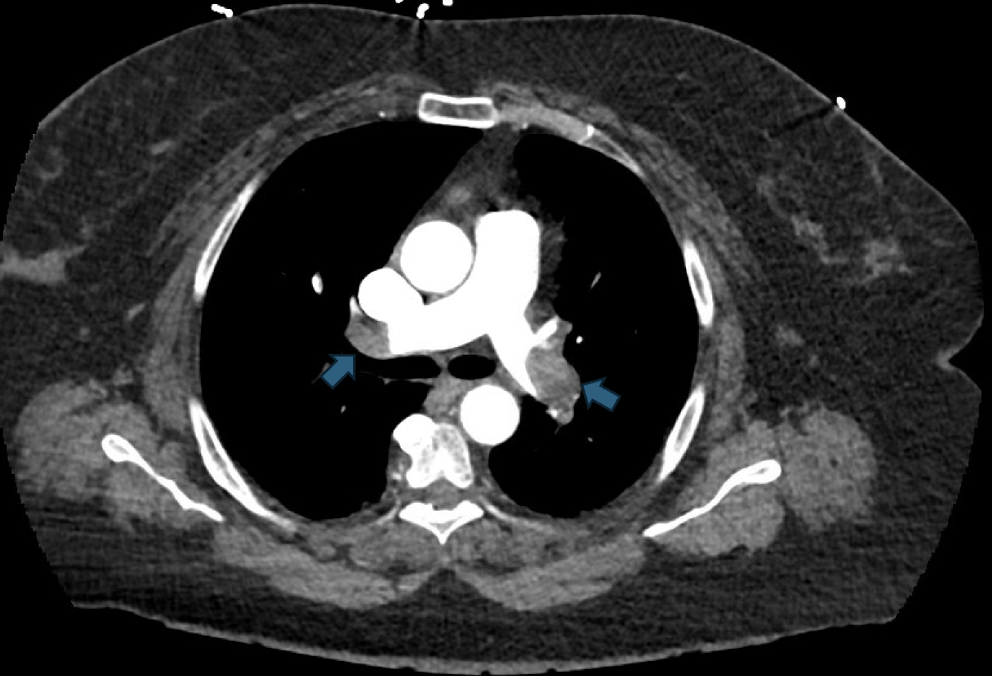
Patient was admitted to the MICU on heparin drip with IR and CT surgery following for possible thrombectomy.
Clinical Pearl:
In patients with high-risk (massive) PE causing hemodynamic instability, acute RV pressure overload can lead to obstructive shock, requiring emergent intervention. Systemic thrombolysis is generally indicated in high-risk PE with persistent hemodynamic instability, provided there are no absolute contraindications. VA-ECMO is an option for massive PE with refractory shock, impending CV collapse or cardiac arrest.