
Patient is a 30-year-old female with PMHx of recurrent pneumonia and Down syndrome presenting to the ED from group home, found to be short of breath and hypoxic to 84% on RA by EMS. VS on arrival shows O2 Sat 96% on 4LNC, RR 20, HR 129, afebrile.
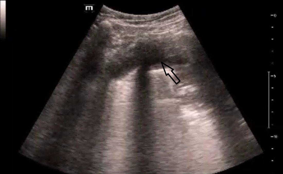
POCUS bedside showed:
In the apical view, you can see lung sliding and the pleural effusion (black arrow):
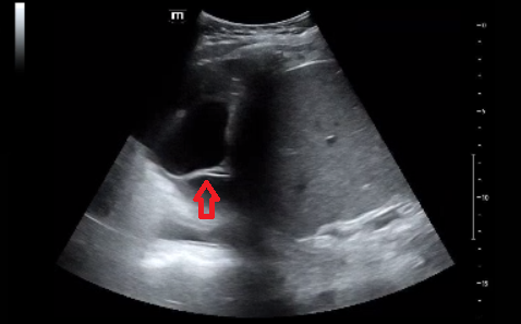
You see a large complex, loculated pleural effusion with mobile septations (red arrow) in the right lung base view.
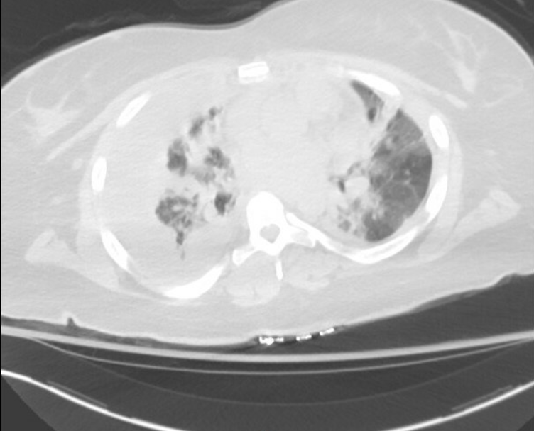
CT chest showed a large pleural effusion measuring higher than water density representing exudate or hemothorax, with nondisplaced sternal and right rib fractures. This is likely a resolving hemothorax in the setting of trauma. CT surgery is consulted and thoracoscopy with pleurodesis was done in the OR revealing hemothorax.
Clinical Pearl: A common cause of loculated pleural effusion is an exudative parapneumonic effusion vs empyema in the setting of pneumonia, requiring IV antibiotics +/- fibrinolysis via chest tube or video-assisted thoracoscopic surgery (VATS) debridement.