POTD: Tinea Pedis (Athlete's Foot)

This PODT is inspired by a recent case I had in while working in Peds and is something we may encounter often in the summer. This is a perfect example of a fast track compliant that we may have not seen a lot of during COVID.

The patient was a young male in his 20s who works in construction and wears heavy boots and socks for about 8 hours of the day in the heat. He presented with 1 days of sloughing of the skin of both of his feet with discharge.

 Lets discuss Tinea Pedis (Athlete’s Foot):

 Tines pedis is a dermatophyte infection of the skin on the foot.

 Etiology and Risk Factors:

  • Usually occurs in adults and adolescents and is rare prior to puberty

  • Infection is acquired by means of direct contact with the causative organism

  • Commonly seen in patients who have a history of walking barefoot in locker rooms or swimming pool facilities

  • Also commonly seen in patients who wear occlusive footwear

Predisposing factors to consider

  • Diabetes Mellitus

  • Immunodeficiency, Systemic corticosteroid use, or use of immune suppressive agents

  • Poor peripheral circulation or lymphoedema

  • Excessive sweating (hyperhidrosis)

 Who would have know that there are different types of tinea pedis?

  •  Types of Tinea Pedis:

    • Interdigital tinea pedis: Manifests as pruritic erosions or scales between the toes, most commonly in the third and fourth digital interspaces

      • More severe form of this is known as Ulcerative tinea pedis. This is generally associated with secondary bacterial infection

    • Hyperkeratotic (Moccasin-Type): Characterized by diffuse hyperkeratotic eruption involving the soles and medial and lateral surfaces of the feet.    

    • Vesiculobullous (inflammatory-type): Pruritic, sometimes painful, vesicular or bullous eruption. Medial foot often affected 

Management:

  • Topical antifungal therapy is treatment of choice for most patients.

    1. Example of topical antifungal: Azoles, Allylamines, Butenafine, Ciclopirox, Tolnaftate, and Amorolfine. Recommended to apply once or twice a day for four weeks. (Refer to references for dosages and frequency)

    2. Beneficial and more effective for patients to use the suspension formulation of these medications

  • Systemic antifungal agents are primarily reserved for patients who fail topical therapy

    1.   Terbinafine 250mg per day for 2 weeks in adults

      1. Most check LFTs prior to administration and patients need to follow up and have LFTs checked while receiving treatment

      2. Peds dosing:

        • 10 to 20kg: 62.5mg/day

        • 20 to 40kg: 125mg/day

        • Above 40kg: standard adult dosing

    • Itraconazole 200mg per day for two weeks

      •   Peds dosing:

        • 3 to 5 mg/kg per day

    • Fluconazole 150mg once weekly for two to six weeks

      • Peds dosing:

        •   6mg/kg once weekly

  • ·Ulcerative Tinea Pedis;

    •   Always treatment with systemic antifungal agents in addition to topical antifungals

    • Make sure to add in addition to your antifungal an antibiotic such as Keflex

    • Outpatient podiatry follow up should be given to patients

  • Prevention

    • Use of sock with wick-away material

    • Use of desiccating foot powders

    • Tx of hyperhidrosis if there is history of moist feet

    •   Tx of shoes with antifungal powder

    •   Avoidance of occlusive foot wear

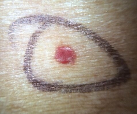
 We diagnosed our patient with ulcerative tinea pedis. We started the patient on Terbinafine, Ciclopriox, and Keflex and arranged for podiatry follow up. Our patients case was unique in the fact that the patient had bilateral involvement normally this occurs unilateral.

 References :

·      https://www.uptodate.com/contents/dermatophyte-tinea-infections?search=tinea%20pedis&source=search_result&selectedTitle=1~103&usage_type=default&display_rank=1#H2658711829

·      https://www.uptodate.com/contents/image?csi=18b425c8-5b1f-4694-a039-5bc8aa27c160&source=contentShare&imageKey=PC%2F76148

·      https://wikem.org/wiki/Tinea_pedis

·      https://www.aafp.org/afp/2014/1115/p702.html

·      https://accessemergencymedicine.mhmedical.com/content.aspx?sectionid=109447903&bookid=1658

·      https://dermnetnz.org/topics/tinea-pedis/

 · 

Everything BURNS!

Good Monday Morning all!

This one took me all weekend - but I think it will be particularly helpful! I am always looking up how to dress burn wounds and who to give followup to and who to transfer etc, so I think this will be a nice easy reference.

There is just too much info around burns (from cyanide poising which we just reviewed w/Dr. Harmouche to awake nasotracheal intubation) and there are just so many learning points and I could only include so many - so I apologize for having the leave some things out. its already a dense one!

Prepare to zoom in/magnify, and happy learning (:

1606701692-2.png

A couple key take home points that I wasnt able to fit into the graphic:

Burns are dynamic wounds. Burns can deepen over the next few days, and so it is difficult to know the true depth of the wound for at least 48-72 hours. Even burn specialists are only correct about 60% of the time at accurately identifying the depth of the burn on initial assessment. Burns can deepen after the first few hours to days of assessment. This is particularly important when setting expectations with patients and families at the initial visit.

Initial approach to any patient in a fire:

Don’t get distracted by the burns. Perform your primary and secondary survey as you normally would with a trauma patient and address the burns later.

Carbon monoxide (CO) and cyanide poisoning are also associated with burn injuries. Apply 100% O2 to reduce the half-life of carboxyhemoglobin to all patients with a history of exposure to fire in an enclosed space. Assume the pt is both trauma AND tox until proven otherwise.

Many of these pictures and pearls came from this amazing website:

https://emergencymedicinecases.com/burn-inhalation-injuries/

 · 

Basics of Dermatology 1

POTD: Basics of Dermatology 1

This pearl will be part of a general overview of our favorite subject dermatology. It is very important to have a basic understanding of describing rashes so that our consultants (who will never come to the ED) can understand what we looking at.

(In fact, I believe dermatology only comes once or twice a week inpatient. I admitted a patient for Stevens-Johnson syndrome that didn't see a dermatologist until 3 days after admission)

Story Time – I will be sharing some of my more memorable cases that I’ve seen in relevant POTDs

This case happened during my intern year during my peds rotation.

13 yo female presenting with fever x 2 days with rash. The patient was in the ED 3-4 days ago with similar symptoms and was discharged as herpangina. The patient’s fever was intermittent (not on contiguous days) and subjective, and there were no other associated symptoms. Vital signs stable in the ED; afebrile in the ED. The patient was well appearing tolerating PO.

On her hands and feet were diffuse macular blanching erythematous undemarcated rash that were non-tender. A few of the lesions were patches about 1 x 1 cm. There were some erythematous lesions in the back of her mouth as well. The patient was discharged with a diagnosis of herpangina.

4 days later (third visit), the patient returned again but now with a bit more fatigue and the rash has gotten a bit worse. The patient was still well appearing but the macules have further progressed into patches over the hands and feet. When we re-examined the rash, underneath the blanching patches, there appeared to be non-blanching petechiae underneath. We got basic labs to rule out Kawasaki’s which were all normal and the patient was admitted; other differential included juvenile idiopathic arthritis and lupus.

The patient turned out to have Lupus.

So thinking back, if I had examined her rash more closely, I might have been able to find the petechial rash on the second visit which would have raised my concern for more systemic pathology. My takeaway is to be more thorough looking through all of the areas of rash as well as a benign patch can be covering a more concerning petechiae.

Basics of the Dermatologic exam

Take a history with OLDCARTS as one normally would with any history.

Choose a primary site and describe its morphology, size, shape, dermarcation, color, distribution. Make sure to look throughout the entire body for other lesions. Often times a patient might have an obvious primary lesion and less obvious ones in flexural surfaces can be missed; an isolated rash can be part of a systemic illness. Any associated symptoms like joint pain, abd pain, n/v/d/c, fevers/chills are key as benign appearing rashes can be a part of a more serious systemic illness.

Definitions

Morphology

Macule: flat, nonpalpable lesion < 0.5 cm                   Ex: Petechiae is a form of a macule

maculenot pete.jpg

Petechiae – small red or brown macules up to 0.5 cm that don’t blanch with pressure

macule.jpg

Patch: flat, nonpalpable lesion > 0.5 cm                      Ex: vitiligo

vitiligo.jpg

Purpura – circumscribed petechiae more than 0.5 cm in diameter

Purpura_vasculitis_arm_0.jpg

Papule: flat, elevated, palpable lesion < 0.5 cm          Ex: cherry angioma, wart

papule.jpg

Plaque: flat, elevated area > 0.5 cm                              Ex. Psoriasis, seborrheic keratosis

plaque.jpg

Nodule: > 2cm dome shaped with a deep component        Ex. Epidermoid cyst

nodule.png

Vesicle: clear fluid filled lesion < 0.5 cm                                 Ex. Herpes, varicella

vesicle.jpg

Pustule: raised fluid filled lesion < 0.5 cm filled with purulent material (similar to vesicle but with pus)          Ex.  Acne

pustule.jpg

Bulla: clear fluid filled lesion > 0.5 cm                                     Ex. Bullous Pemphigoid, blister

bullous pemphigoid.jpg

Size

Shape

Round/discoid coin shaped, no central clearing (nummular eczema)

Oval ovoid pityriasis rosea Annular round, with active margin and central clearing (tinea corporis) Reticular net-like or lacy (lichen planus)

Linear in a line contact dermatitis Iris/target purple papule in the center of pink macule (erythema multiforme)

Serpiginous snakelike or wavy line track (cutaneous larval migrans)

Polycyclic interlocking or coalesced circles (psoriasis)

Morbilliform measles-like; maculo-papular lesions with confluence on the face and body (roseola, mononucleosis)

Demarcation: are the borders clearly defined? erysipelas vs cellulitis

Color: erythematous (cellulitis, drug reaction), loss of pigmentation (vitiligo), violaceous (Kaposi sarcoma), brown, yellow (xanthoma), black (eschar), honey-colored (impetigo)

Secondary morphology: serum/dried crust (impetigo), fissure, lichenification (thickening of skin), erosion (partial loss of epidermis), ulceration (full thickness loss of epidermis), skin sloughing

Distribution: extensor surface (psoriasis), flexor (atopic dermatitis), generalized (drug eruption), underneath belt/watch (contact dermatitis), dermatomal (varicella)

Dermatology is hard and there is an entire residency for it so we will never know everything about dermatology but there are a few can’t miss dermatologic emergencies that we can’t miss that will be covered in the next POTD!

- Kevin