History:
Symptoms include SOB and chest pain.
Remember this may manifest as back pain depending on mechanism.
Look for in high impact injuries to chest (MVC, fall, pedestrian struck, trampled by livestock, etc)
MOA being compression-decompression.
Exam:
Flail chest or crackles (however unlikely unable to auscultate in ED).
Observe for crepitus for possible pneumothorax.
Seatbelt sign.
Diagnosis:
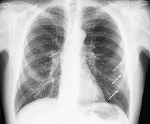
CXR or CT chest
Extent of injury not apparent on CXR for 24-48 hours
Areas of lung opacification within 6 hours diagnostic of pulmonary contusion.
There are NEXUS chest guidelines (yes, chest!) for patients>14 to omit any imaging in chest trauma (see appendix below) - 98.8% sensitive.
Look for homogenous focal or diffuse opacity that may cross typical anatomical landmarks (i.e. lobes).
Treatment:
Primarily supportive. Watch for delayed presentation!
Consider Bipap; pain control with intercostal block or epidural inpatient. Avoid unnecessary fluids.
Up to 40-60% will require mechanical ventilation. Also may be necessary to sedate for pain control.
Place good lung in dependent position to improve V/Q mismatch 50% go on to develop ARDS (blood in alveoli activates inflammatory cascade).
If not improving - ECMO (V-V) is a possibility.
Bottom line:
Monitor patients suspicious for pulmonary contusion - if they have signs of CXR there is a good chance they may need more invasive support (e.g. intubation).
Have low suspicion for concurrent injuries including mediastinal and vascular injuries, diaphragmatic rupture, and cardiac contusion.
Be aware of patient fluid status and try not to overload patient.

Keywords: Pulmonary Contusion NEXUS Chest Radiography Chest Trauma