Resuscitative TEE

Important: Please complete the Sexual Harassment Online Module ASAP!


Resuscitative Transesophageal echocardiography (TEE)

  • TEE allows the emergency physician to maintain the standard of an ultrasound-informed resuscitation in the scenario of cardiac arrest, where TTE is significantly limited.

  • Focused or resuscitative TEE (4 views) differ from comprehensive TEE (>20 views) that cardiology performs in that it is employed to identify specific questions.

  • TEE allows for potentially shorter chest compression pauses

  • TEE allows for evaluation for the quality of chest compressions

  • TEE allows for visualization of fine V-fib not seen on the monitor


Indications: Cardiac arrest (ACEP)

Contraindications: Esophageal injury or stricture and lack of a definitive airway

How to manipulate a TEE Probe:

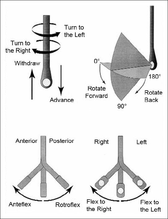
5 different ways you can physically manipulate the TEE probe

1. Withdraw or Advance up or down patient’s esophagus

2. Turn probe to right or left

3. Turn tip of flip in anterior- ante-flexing or in the posterior direction called retro-flexing --> large wheel

4. Turn tip to Left or right -->  small wheel (not typically used for our purposes)

5. In addition, you can rotate the transducer housed within the probe itself (AKA omniplane or multiplane)-->  adjusts the beam angle anywhere between 0° and 180° -->  two smaller buttons ( crystal rotation)

TEE manipulation.jpg


TEE-controls- wheels.png


The views are obtained in the following order: : 

The midesophageal 4-chamber view (ME 4C) is obtained by advancing the TEE probe to the thoracic esophagus and orienting the multiplane at 0-20° in neutral flexion. You may need to retroflex slightly to see all four chambers.

-   The midesophageal long-axis view (ME LAX) is obtained by leaving the probe in the same location as the midesophageal 4-chamber, but increasing the multiplane to between 110° and 160° while in neutral flexion. 

-   The transgastric short axis view (TG- SAX) is obtained by first moving the multiplane to 0°, then advancing the probe into the stomach and ante-flexing the probe

-   The bicaval view (ME bicaval) is obtained by turning the entire probe to the patient’s right towards the superior vena cava (SVC) and inferior vena cava (IVC) while in the mid-esophagus, keeping the multiplane at 90-100° with neutral flexion

 ( The first 3 views are recommended by ACEP. Bicaval not recommended by ACEP) 


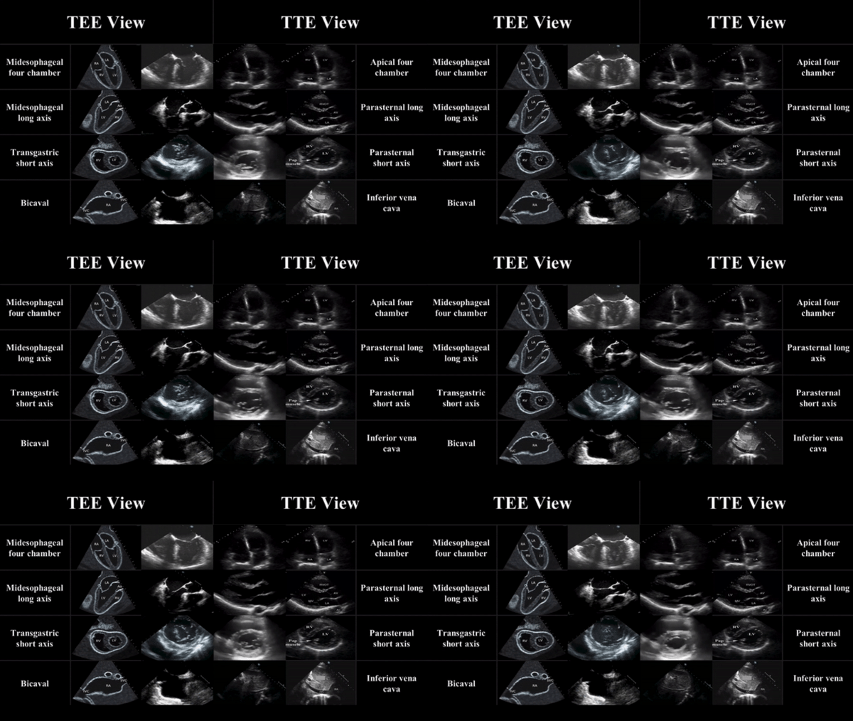
TEE views and their analogous TTE views


TTE-and-TEE.gif

Midesophageal four chamber view (ME 4C)

-  Apical four chamber view

-  Great visualization of all chambers as well as the tricuspid and mitral valves in one plane.

-   Evaluation of right and left ventricular systolic function and size

-   Preferred view to evaluate for the presence or absence of a perfusing rhythm during a pulse check.

ME4C.png


Midesophageal Aortic Long Axis view (ME LAX)

-  Midesophageal analogous to the parasternal long axis view in TTE

-   View includes the mitral and aortic valves, as well as the left atrium, left ventricle, and left ventricular outflow tract of the right ventricle.

-   Evaluate left ventricular systolic function, and provides feedback on compression adequacy and location. High-quality compressions cause maximal compression of the left ventricle and visualization of the aortic valve opening and closing indicating forward flow of blood.  Poor quality compressions are seen over the aortic root and there is no valvular indication of forward flow. 

ME- LAX.png


Transgastric Short Axis view (TG- SAX)

- Analogous to the parasternal short axis TTE view

- Evaluate left ventricular systolic function, including any regional wall motion abnormalities

- Can evaluate for acute MI and the presence of septal flattening in this view

TGSAX.png

Mid Esophageal Bicaval View (ME bicaval)

-   Analogous to the inferior vena cava view of TTE

-   Transducer plane cuts through the left atrium (LA), right atrium (RA), IVC and SVC.

This view allows the operator to evaluate for hypovolemia, atrial size, and interatrial septum bowing.

-   Aids in the placement of central venous catheters, transvenous pacemakers, or extracorporeal life support (ECMO) vascular cannulas by observing the initial wire placement in the vasculature

- Can aid inevaluation of fluid status to guide fluid resuscitation (looking at respiratory variation in SVC)

bicaval.png


Pitfalls

-  Compressions do not need to be stopped for TEE insertion. Additionally, the TEE can be left in the esophagus during defibrillation. The probe should be inserted or withdrawn while the tip is in neutral position, and not while the tip is flexed to avoid esophageal injury. 

-  Images should be optimized to avoid foreshortening of the ventricles and to include the appropriate structures for each view.

-  Pericardial effusions must be taken into clinical context, as small effusions can cause tamponade if accumulated rapidly, while large effusions can be well tolerated if they accumulate slowly.

-   Clotted hemopericardium may be isoechoic with the myocardium, making it difficult to identify.

-  Right ventricular failure is not specific to pulmonary embolism, and can be due to pulmonary hypertension or other etiologies such as right sided myocardial infarction, or even cardiac arrest itself.

-  Pleural effusions can be mistaken for pericardial effusions. Multiple views should be used to corroborate findings.

-  Fat pads can be mistaken for pericardial effusions, but these are hypoechoic rather than anechoic and limited to the anterior and apical regions of the heart, not circumferential.


 Resource: 

Check out this 3D module that you can practice on 

https://pie.med.utoronto.ca/TEE/TEE_content/TEE_standardViews_intro.html

References:

Drs Lawrence Haines, Judy Lin and Alyssa Phuoc-Ngyuyen

Images: Adapted from Arntfield R, Pace J, McLeod S, et al. Focused transesophageal echocardiography for emergency physicians-description and results from simulation training of a structured four-view examination. Crit Ultrasound J. 2015;7(1):27.

Teran, Felipe, et al. "Evaluation of out-of-hospital cardiac arrest using transesophageal echocardiography in the emergency department." Resuscitation 137 (2019): 140-147.

EmDocs

ACEP policy statement

https://www.acep.org/patient-care/policy-statements/guidelines-for-the-use-of-transesophageal-echocardiography-tee-in-the-ed-for-cardiac-arrest/

 · 

“Pressors” in Distributive Shock in Adults

“Pressors” in Distributive Shock in Adults

Thank you, Dr Dastmalchi, for requesting this POTD. I will review “pressors” for cardiogenic shock separately

  • Vasopressors- Pure vasoconstriction without any inotropy eg Phenylephrine and Vasopressin

  • Inotrope- Increase cardiac contractility à improving SV and cardiac output without any vasoconstriction eg Milrinone

  • Inopressors - a combination of vasopressors and inotropes, because they lead to both increased cardiac contractility and increased peripheral vasoconstriction eg Norepinephrine, Epinephrine and Dopamine

Norepinephrine- Inopressor

  • First line vasopressor in septic shock per Surviving Sepsis Guidelines

  • Less arrhythmogenic than Epinephrine and Dopamine


Mechanism of action

  • Stimulates alpha-1 and alpha-2 receptors

  • Small amount of beta-1 agonist- modest inotropic effect

  • Increased coronary blood flow and afterload

  • Increases venous tone and return with resultant increased preload

Adverse effects

  • Norepinephrine is considered safer than both Epinephrine and Dopamine.

  • ARR of 11% compared to dopamine with NNT 9

  • NE superior in improving CVP, urinary output, and arterial lactate levels compared to Epinephrine, Phenylephrine, and Vasopressin.

Indications

  • First-line pressor choice in distributive shock, including both neurogenic and septic shock

  • Norepinephrine as the only first-line pressor per SSC guidelines

Dosing

  • Use weight-based dosing to avoid the adverse effects associated with norepinephrine use

  • Weight-based dosing is based on GFR

  • Norepinephrine has a rapid onset of action (minutes) and can be titrated every 2-5 minutes

Epinephrine- Inopressor

Mechanism of action

  • Beta-1 and beta-2 receptors agonism à more inotropic effects than norepinephrine

  • Epinephrine greatly increases chronotropy (heart rate) and thus stroke volume

  • Some stimulatory effect on alpha-1 receptors

  • Lower doses (1-10 mcg/min) à a beta-1 agonist

  • Higher doses (greater than 10 mcg/min) à an alpha-1 agonist

Adverse effects

  • Associated with an increased risk of tachycardia and lactic acidosis

  • Hyperglycemia

  • Increased incidence of arrhythmogenic events associated with epinephrine

  • More difficult use lactate as a marker of the patient’s response to treatment

Indications

  • SSC guidelines recommend epinephrine as a second-line agent, after norepinephrine

  • “push-dose pressor”

  • Due to beta-2 receptors agonism causing bronchodilation, epinephrine is first-line agent for anaphylactic shock

Dosing

  • Guidelines for anaphylactic shock recommend an initial bolus of 0.1 mg (1:10,000) over 5 minutes, followed by an infusion of 2-15 mcg/min however associated with adverse cardiovascular events

  • For septic shock start epinephrine at 0.05 mcg/kg/min (generally 3-5 mcg/min) and titrate by 0.05 to 0.2 mcg/kg/min every 10 minutes. maximum drip rate is 2 mcg/kg/min

Dopamine- Inopressor

  • Fallen out of favor

  • Associated with higher arrhythmogenic events

Mechanism of action

  • Effects are dose-dependent

  • Low doses à dopaminergic receptors à leads to renal vasodilation à increased renal blood flow and GFR although studies failed to demonstrate improved renal function with dopamine use clinically

  • Moderate doses à beta-1 agonism à increased cardiac contractility and heart rate

  • High doses à alpha-1 adrenergic effects à arterial vasoconstriction and increased blood pressure

Adverse Effects

  • Several large, multi-center studies that demonstrate increased morbidity associated with its use

  • Significantly higher rates of dysrhythmias à NNH 9

Indications

  • Rescue medication when shock is refractory to other medications

Dosing

  • Start at 2 mcg/kg/min and titrate to a maximum dose of 20 mcg/kg/min.

  • Less than < 5 mcg/kg/min à vasodilation in the renal vasculature

  • 5-10mcg/kg/min à beta-1 agonism

  • 10 mcg/kg/min à alpha-1 adrenergic

Vasopressin- Vasopressor

  • Add vasopressin (doses up to 0.04 units/min) to norepinephrine to help achieve MAP target or decrease norepinephrine dosage

  • Restore catecholamine receptor responsiveness, particularly in cases of severe metabolic acidosis.

  • pH independent

  • Pure pressor à increase vasoconstriction with minimal effects on chronotropy or ionotropy

Mechanism of action

  • At low doses (< 0.04 units/min) à increases vascular resistance (V1)

  • No effect on heart rate and cardiac contractility

Adverse Effects

  • Vasopressin has been shown to be as safe as norepinephrine at lower doses

  • Increases SVR and afterload and decreases cardiac output although unclear if effect significant at lower doses

Indications

  • Second line vasopressors per SSC guidelines for septic shock

  • pH independent- Vasopressin in combination with epinephrine demonstrated improved ROSC in cardiac arrest patients with initial arterial pH <7.2 compared with epinephrine alone

Dosing

  • Steady dose at 0.03-0.04 units/min

  • Vasopressin is not titrated to clinical effect as are other vasopressors

  • Think about it more as a replacement therapy and treatment of relative vasopressin deficiency

Phenylephrine- Vasopressor

  • Pure pressor à increase vasoconstriction with minimal effects on chronotropy or ionotropy

  • SSC guidelines does not make rated recommendations on Phenylephrine

  • Limited clinical trial data

Mechanism of action

α1 agonism with peripheral vasoconstriction

Adverse Effects

  • Bradycardia - decrease in heart rate mediated by the carotid baroreceptor reflex 2/2 increase in SVR

  • Increases SVR and afterload and decreases cardiac output

Indications

  • Patients that are susceptible to beta-adrenergic generated arrhythmia

  • Push dose formulation

  • Refractory shock

Dosing

0.1-2mcg/kg/min (onset: minutes, duration: up to ~20 minutes)

References:

Emdocs

LITFL

Pollard, Sacha, Stephanie B. Edwin, and Cesar Alaniz. "Vasopressor and inotropic management of patients with septic shock." Pharmacy and Therapeutics 40.7 (2015): 438.

Amlal, Hassane, Sulaiman Sheriff, and Manoocher Soleimani. "Upregulation of collecting duct aquaporin-2 by metabolic acidosis: role of vasopressin." American Journal of Physiology-Cell Physiology 286.5 (2004): C1019-C1030.

Khanna, Ashish, and Nicholas A. Peters. "The Vasopressor Toolbox for Defending Blood Pressure."

Turner, DeAnna W., Rebecca L. Attridge, and Darrel W. Hughes. "Vasopressin associated with an increase in return of spontaneous circulation in acidotic cardiopulmonary arrest patients." Annals of Pharmacotherapy 48.8 (2014): 986-991.

 · 

Targeted Temperature Management

Great job on resuscitating that V fib cardiac arrest and achieving sustained ROSC. 

Now what? Cool them!

Whether you cool them or not could determine whether your patient goes into multisystem organ failure in the ICU or walks out of the hospital few weeks later.


What:

Targeted temperature management (TTM) to improve survival and neurological outcomes among comatose survivors of patients with cardiac arrest


Who:

Adults with out-of-hospital cardiac arrest with an initial shockable rhythm and nonshockable rhythm


Inclusion criteria (must meet all criteria)

  • Postcardiac arrest status (any rhythm as a cause of arrest is eligible)

  • ROSC < 30 minutes from EMS/code team arrival

  • Time at induction < 6 hours from ROSC

  • Comatose status (patient does not follow commands)

  • MAP ≥ 65 mm Hg (may include use of vasopressor drugs)

Exclusions may include

  • DNR advanced directive, MOLST, poor baseline status, or terminal disease

  • Traumatic etiology for the arrest

  • Active bleeding or known intracranial bleeding (relative)

  • Cryoglobulinemia (relative)

  • Pregnancy (relative; consider obstetrician/gynecologist consultation)

  • Recent major surgical procedure (relative)

  • Severe sepsis/septic shock as cause of arrest (relative)

Why:

  • Decreased fever-related tissue injury

  • Reduction in ischemic-reperfusion injury

  • Cerebral metabolic rate decreases by a 6-7% for every 1ºC drop in body temperature which reducing oxygen demand, preserving phosphate compounds and preventing lactate production and acidosis

  • Bernard, et al (2002) found an Absolute Risk Reduction (ARR) for death or severe disability of 23%, NNT was 4.5

  • The Hypothermia After Cardiac Arrest (HACA) Group (2002) found an ARR for unfavourable neurological outcome of 24%, and NNT of 4


How:

  • IV cold saline 2-3 mL/kg

  • Cooling vest and cooling machine- Arctic Sun

  • If shivering does not occur, do not use neuromuscular blockade

  • If paralysis employed, titrate to degree of shivering- do not need train-of-four monitoring

  • Sedation of choice is institution dependent (MMC CICU uses Fentanyl and Midazolam)

When:

Initiation of TTM within122 minutesof hospital admission was associated with improved survival.
Most guidelines recommend initiation within6 hours

What temperature should be targeted:

This remains controversial, with guidelines accepting a range of temperature targets from 33-36C. Available evidence shows no benefit to hypothermia (33C) compared to normothermia (36C). In the absence of evidence, targeting 36C is prudent

  • TTM36 is more hemodynamically stable than TTM33, which is relevant because these are often very unstable patients.

  • TTM36 avoids electrolytic shifts associated with raising and lowering the temperature.

  • Hypothermia at 33C suppresses immune function and associates with increased rates of pneumonia.

  • TM33 will induce bradycardia, which is dangerous in patients with underlying torsades de pointes.

References

Bernard SA et al. Treatment of comatose survivors of out-of-hospital cardiac arrest with induced hypothermia. N Engl J Med 2002;346:557-63. PMID 11856794

Hypothermia after Cardiac Arrest Study Group. Mild therapeutic hypothermia to improve the neurologic outcome after cardiac arrest. N Engl J Med 2002;346:549-56. PMID 11856793

Nielsen N et al. Targeted temperature management at 33 degrees C versus 36 degrees C after cardiac arrest. N Engl J Med 2013; 369: 2197-206. PMID 24237006

Stanger, Dylan, et al. "Door‐to‐targeted temperature management initiation time and outcomes in out‐of‐hospital cardiac arrest: insights from the Continuous Chest Compressions Trial." Journal of the American Heart Association 8.9 (2019): e012001.

Donnino, Michael W., et al. "Temperature management after cardiac arrest: an advisory statement by the advanced life support task force of the international liaison committee on resuscitation and the American Heart Association emergency cardiovascular care committee and the council on cardiopulmonary, critical care, Perioperative and Resuscitation." Circulation 132.25 (2015): 2448-2456.

REBEL EM
LITFL
EB Medicine
Mayo Clinic Florida TTM Guideline

 ·