Pelvic Inflammatory Disease & Tubo Ovarian Abscess

Pelvic inflammatory disease (PID) is an infection of the upper reproductive tract, which includes the uterus, fallopian tubes, and ovaries. The most common pathogens are gonorrhea and/or chlamydia which begin as a cervical infection and become polymicrobial as they ascend. Symptoms may include fever, nausea, vomiting, malaise, abdominal pain, purulent vaginal discharge, or abnormal vaginal bleeding. Unilateral adnexal tenderness/fullness may indicate a developing tubo ovarian abscess (TOA), a complication of PID. 

A TOA is an inflammatory mass that involves the fallopian tube, ovary, and sometimes other adjacent pelvic organs (bladder, bowel). They may require aggressive medical and/or surgical therapy as a ruptured TOA can result in sepsis. Treatment ranges from antibiotics to laparoscopy. Some stable, non-ruptured TOAs can be treated with antibiotics alone. Suggested antibiotic regimens include: 

  • CTX 1g qd + doxycycline 100mg q12 + metronidazole 500 mg q12

  • Cefotetan 2g IV q12 + doxycycline 100mg q12

  • Cefoxitin 2g IV q6 + doxycycline 100mg q12.

Studies suggest that abscesses =/> 7 cm have a higher likelihood of requiring surgical therapy (drainage or surgical removal). Therefore, it is appropriate to trial IV abx if the patient is hemodynamically stable, has adequate response to initial IV abx, and has imaging that shows that the abscess is < 7 cm. 

Diagnosis can be made via transvaginal US or CT A/P. Conventional teaching is that US is the preferred modality for imaging pelvic organs to assess for TOAs. However, recent studies have shown that CT has a higher sensitivity for diagnosing TOAs. Therefore, common practice is to start with US as it helps rule out other pathology, such as ovarian torsion, and is less expensive and less radiation for the patient. A positive US can help establish the diagnosis, however, a negative US does not exclude a TOA and a CT is often indicated. Ultimately, TOAs are a clinical diagnosis and are often diagnosed in the setting of pelvic mass in patients who meet the diagnostic criteria for PID. These patients should get an OBGYN consult and be started on IV abx. 

Thanks for reading!

Ariella

Resources: 

  1. Fouks Y, Cohen A, Shapira U, et al. Surgical Intervention in Patients with Tubo-Ovarian Abscess: Clinical Predictors and a Simple Risk Score. J Minim Invasive Gynecol 2019; 26:535

  2. Lee"DC,"et"al.)Sensitivity)of)ultrasound)for)the)diagnosis)of)tuboAovarian)abscess:)A)case)report)and) literature)review.))J(Emerg(Med."2010"May"11"

  3. https://www.uptodate.com/contents/epidemiology-clinical-manifestations-and-diagnosis-of-tubo-ovarian-abscess?search=tuboovarian%20abscess&topicRef=16419&source=see_link

  4. https://www.uptodate.com/contents/management-and-complications-of-tubo-ovarian-abscess?search=tuboovarian%20abscess&source=search_result&selectedTitle=1~21&usage_type=default&display_rank=1


Acute Compartment Syndrome

Acute compartment syndrome is when the pressure in a muscle compartment increases, compromising circulation and function. This occurs because the compartment is surrounded by a fascial membrane that restricts further expansion. It typically occurs after trauma, crush injury, or burns. Signs include severe pain (earliest sign), pallor, paresthesia, paresis, and pulse deficit. To measure compartments, you take your measurement device and insert it into the compartment of interest. 

How to set up your measurement device: 

  • Your materials include a sterile 3cc saline syringe, chamber, and needle. The needle has a side port (hole) for measuring pressure.

  • Connect the syringe, chamber, and needle

  • Flush the chamber and needle with saline to get rid of the air; do this by holding the entire device at a 45-degree angle.

  • Load into the monitoring unit and press zero, you should see 00

  • Insert the needle into the compartment of choice and hold it for reading

There are two ways to assess for compartment syndrome. You can use the absolute or delta pressure (normal: 0-8). Suspect compartment syndrome if:

  • the absolute pressure is > 30 mmHg

                                   OR

  • The delta pressure is < 30 mmHg

    • Delta pressure = diastolic pressure - compartment pressure. This means that the pressure in your compartment is so high that it is close to your diastolic blood pressure

Tip: remember the number 30 

These patients require a fasciotomy so call ortho ASAP. Meanwhile, you should level their affected limb and support BP if hypotensive to help maintain perfusion. 

Thanks for reading!

-Ariella

References: 

https://www.emrap.org/episode/trauma1/compartment

https://www.emrap.org/episode/measuring/measuring


POTD: Umbilical Vein Catheterization

I wanted to review a fairly rare but lifesaving EM procedure in neonates. This procedure is done fairly commonly in the NICU/L&D, but is done less frequently in the ED, especially with our excellent nurses who can literally get the most impossible venous accesses. If you went to Airway day, you might recall Dr. Sokolovsky describing her harrowing tale of providing neonatal resuscitation at Burning Man and performing an umbilical vein catheterization with an 18-gauge IV. Super wild! So for anyone who might find themselves in a similar poop-inducing situation with no pediatric support or NICU available, this is for you!


Umbilical vein catheterization is indicated in a neonate within 14 days post-birth requiring IV resuscitation. The stump must be "fresh", so it is most ideal in the newly born neonate. Here is an excellent video overviewing the following steps. https://pedemmorsels.com/wp-content/uploads/2019/08/UVC....mp4

Here's what you'll need:

  • Sterile gloves (gown and drape less non urgent)

  • chlorhexidine

  • forceps

  • scalpel

  • umbilical line (5 French is standard, 3.5 French in very premature baby)

  • three-way stopcock

  • umbilical tape of 3-0 silk/nylon

  • NS flush

In peds, we have umbilical vein catheterization trays located on the top shelf in Bay 31 that includes all of the above except the catheter. While the umbilical line is the traditional teaching, you can use any tube that can fit into the vein - that means an 18 gauge IV, pediatric central line, feeding tube, etc


Prep the umbilical stump

  1. Flush the line and place

  2. Sterilize the entire umbilical stump, including the clamp at the end of the stump, and the abdomen

  3. Tie the umbilical tape (or a silk string) around the base of the stump loosely. This helps decreased blood flow for when the clamp is eventually removed. It can also be tightened to secure the line once placed

  4. Holding the clamp, make a transverse cut off the stump to remove the distal tip. Cut should be made directly below the clamp or 2 cm from the abdomen.

Identifying umbilical vein and prep for insertion

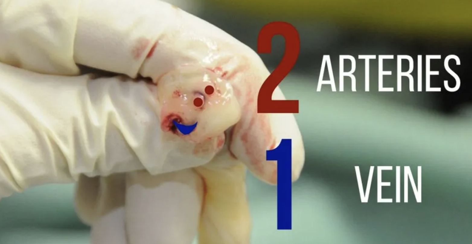
  1. Identify the umbilical vein. The anatomy of the stump involves two smaller umbilical arteries and one umbilical vein. The arteries are typically smaller and thicker lumen, while the vein is larger and more collapsible (see below)

  2. Remove any clots from the vein and gently dilate the vein with forceps

  3. gently insert the line, when you get blood return insert 1-2 cm deeper, or approx 3-5 cm. If there is resistance, consider loosing the umbilical string.

  4. Aspirate blood and flush with NS. Secure the line by tightening the umbilical string and securing with tape or purse string suture

Complications of UVC placement are similar to CVP placement: excessive bleeding, infection, thrombosis, arterial insertion. Specifically to UVC is risk of insertion too deep into the portal venous system or right atrium, which can lead to hepatic necrosis and perforation.

Resources:

https://first10em.com/umbilical-vein-catheterization/

https://wikem.org/wiki/Umbilical_vein_catheterization

https://www.ncbi.nlm.nih.gov/books/NBK549869/