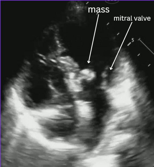
VOTW: Mass-ive Fever

This weeks’ VOTW was brought to you by Drs. Hannah Blakely, Patricia Camino, and the ultrasound team that was scanning that day!

HPI: 6 year old female with PMH of atrioventricular canal defect s/p repair, recent strep throat infection presenting for fever x 14 days.

Bedside POCUS showed:

Evaluating for endocarditis on POCUS:

B mode: look for masses, usually on the lower flow side of the valves (ex: mitral valve- endocarditis is more likely to be found on atrial side).

Color flow: you can usually find associated regurgitation of the affected valve

Possible mimics:

  • Thrombus

  • Papillary muscle rupture/flail leaflet

  • Intracardiac tumor

  • Artifact

Remember that a valve vegetation is one of the major diagnostic criteria for endocarditis. In the right clinical scenario this POCUS finding can highly increase your suspicion for endocarditis.

Case conclusion: CTAP significant for possible splenic and renal infarcts. Patient was admitted for suspected endocarditis. Blood cultures were +MSSA. Pediatric cardiology ECHO was consistent with mitral valve vegetation consistent with endocarditis and septic emboli.

Resources for more info:


Happy scanning!

  • The US Team


VOTW: You take my breath away!

HPI: 90 yo female presenting for worsening shortness of breath and tachycardia x 3 days and right leg pain x 2 weeks with difficulty ambulating.

POCUS showed:

ECHO A4C view (see video): note the size of the RV appears larger than the LV. This is a sign of right heart strain and in the appropriate setting (such as this one) can be concerning for a pulmonary embolism!

Compression views of the common femoral vein (CFV), femoral vein (FV), and popliteal vein (PV). See the echogenic material inside the popliteal vein which is suggestive of a DVT. Remember that during the acute phase of a DVT (<14 days), the clot may appear isoechoic to the blood inside the vein so you may not see this echogenic material and should rely more on your compression exam.

Review on how to do DVT US:

Linear probe

Patient in frog leg position

4 main areas to view

  • Common femoral vein (CFV)-saphenous vein junction (SFV)

    • Clot noted in the SFV within 3 cm to the junction is treated as a DVT. More distally, if there is 5 cm worth of clot noted in the SFV it is also treated as a DVT.

  • CFV branching into [superficial] femoral vein and deep femoral vein

  • Mid/distal femoral vein

  • Popliteal vein

    • Remember the popliteal vein is on top of the popliteal artery (pop on top!)


Tips:

  • You often have to go much higher in the groin than you think to find the CFV-SFV junction

  • Compression testing of the deep veins should not compress the artery (if it is, you’re pressing too hard and can miss subtle DVTs)

  • Deep veins are paired with arteries so identify your landmarks to ensure you are looking at the correct vessels

  • Use your non-scanning hand to help with compression of deeper veins by supporting the other side of the patient’s leg

Case conclusion: Patient with elevated troponin and BNP. CTA significant for bilateral PE’s. Labs and ECHO findings consistent with submassive PE. Patient started on heparin drip and admitted to the floor!

Happy scanning!

  • The US Team


POTD: Hematuria

This POTD was requested by one of our star interns and maybe my long lost distant relative, Dr. Alan Chung!

Today, we're talking about hematuria and our approach as EM providers. 


What is it?

Hematuria is defined as >3-5 RBCs per HPF on urine microscopy. Gross hematuria means that, by just looking at the urine, it looks bloody. Just >1ml of blood in the urine can cause gross hematuria.


Causes of hematuria


This is a long and exhaustive list, but common causes we see in the ED include UTIs (cystitis, pyelonephritis), nephrolithiasis, trauma from catheterization, and blunt or penetrating trauma. Also, don't forget to ask your female patients if they are on their menstrual period.

However, can't miss diagnoses include AAA, renal artery embolus/infarct/dissection, renal vein thrombosis. And of course, if the suspicion for malignancy is high, you want your patient to be informed of this possibility and have close follow up. 


Is it really hematuria?

Pseudohematuria, or urine that appears grossly bloody but on urine microscopy actually has no RBCs, can be caused from a variety of reasons. Common ones include rhabdomyolysis, medications such as nitrofurantoin, pyridium, and rifampin, and foods such as beets and artificial food colorings. So, before you start really thinking about what's causing the hematuria, make sure you get a UA and confirm that it is, in fact, blood. 


Work up

Asymptomatic microscopic hematuria can oftentimes be benign, in the absence of any significant risk factors. Oftentimes, if these patients are stable, asymptomatic, and does not have significant risk factors, they can follow up with their primary care doctor.

Painless gross hematuria is more concerning for malignancy, and would benefit from close urologic follow-up. Other risk factors for malignancy include older age, smoking, family hx, hx of occupational exposure. 

Ask about urinary retention - if the pt is passing clots, this may obstruct the urethra, leading to a lower urinary tract obstruction.


Otherwise, if your patient's history, signs/symptoms, or exam points you to another diagnosis (such as nephrolithiasis, vascular disease, nephropathy...etc), you may want to obtain additional labs and imaging. 


Treatment

Once again...depends! As noted above, stable patients with asymptomatic microscopic hematuria can oftentimes follow up with their PCP. Patients with painless gross hematuria but no urinary obstruction, no AKI, and is otherwise stable should be referred for close urology follow up.

If you patient is unable to urinate, has a significant decline in renal function, decline in Hgb/Hct, or of course if you find any of the big bad vascular causes of hematuria, the patient should be admitted. 


Special considerations for pediatrics

In kids, pay special attention to post-infectious glomerulonephritis and Henoch-Schonlein Purpura

Gross hematuria with edema, proteinuria, and/or hypertension? -> think renal causes like nephritic/nephrotic syndromes, oftentimes post-infectious glomerulonephritis 

Gross hematuria with abdominal pain and rash? -> think HSP


References

Willis GC, Tewelde SZ. The Approach to the Patient with Hematuria. Emerg Med Clin North Am. 2019;37(4):755-769. doi:10.1016/j.emc.2019.07.011

https://www.emdocs.net/em3am-hematuria/

https://www.emdocs.net/evaluation-and-management-of-hematuria-in-the-emergency-department/

https://www.emra.org/emresident/article/hematuria-management

https://pedemmorsels.com/microscopic-hematuria/

https://www.auanet.org/meetings-and-education/for-medical-students/medical-students-curriculum/urologic-emergencies

https://www.ebmedicine.net/topics/hepatic-renal-genitourinary/pediatric-hematuria

 ·