Blood is thicker than water

Hello all, this week's video of the week is a spooky bloody one inspired by this past Halloween weekend! 

Brought to you by Dr. Gabriela Hernandez and Dr. Victor Wong!

ED Course

61 y/o female with PMHx of HLD presents to the ED for worsening suprapubic pain x5 months. Several months ago she had an MRI which revealed free fluid in her pelvis.  States that she had an outpatient ultrasound done 4 days ago which revealed persistent fluid in the pelvis and she was sent home with pain medication and antibiotics. Associated symptoms are subjective fever, pelvic pain with walking, and dysuria. Reports history of C-section x1 and D&C. Denies nausea, vomiting, diarrhea, chest pain, vaginal bleeding, vaginal discharge.

Ultrasound

In this still image from clip 1, there is some free fluid around a distended uterus that is filled with heterogenic complex fluid that may be a mass or coagulated blood.

In the 2 attached clips, clip 1 shows a sagittal distended uterus with complex fluid, likely blood clots with trace amount of free fluid in the pelvis. Clip 2 shows the transverse view of the uterus again with complex fluid.

Formal ultrasound obtained by the team revealed: Markedly distended uterus containing complex fluid. Considerations include endometrial carcinoma vs. extensive blood clot. Possibly infection/pyometrium in the appropriate clinical setting.

Conclusion:

OBGYN consulted. They recommended obtaining tumor markers and close follow-up with her outpatient GYN.

Learning Points:

Appearance of blood on ultrasound can be variable depending on age of clot

  • Acute, fresh blood

    • Often hypoechoic or anechoic, may resemble simple fluid early on

    • Can have internal echoes that swirl with probe pressure

  • Organizing / clotted blood:

    • Becomes heterogeneous, with low-to-medium echogenicity

    • Often appears as avascular, nonshadowing, ill-defined material

    • Can seem like a “soft tissue–like” mass that lacks internal vascularity on color Doppler

    • Echogenicity tends to increase as the clot ages and fibrin organizes

  • Chronic clot:

    • Can become retracted and hyperechoic, sometimes mimicking a fibroid or retained products

References:

Patel, S. J., Feldstein, V. A., & Filly, R. A. (2021). Sonographic differentiation of retained products of conception, blood clot, and intrauterine masses: Diagnostic challenges and clinical implications. Emergency Radiology, 28(3), 527–534. https://doi.org/10.1007/s10140-020-01865-6

Radiopaedia contributors. (2025). Blood clot (ultrasound). Radiopaedia.org. Retrieved from https://radiopaedia.org/articles/blood-clot-ultrasound

Nassiri, S., & Lerman, J. (2017). Ultrasound features of pelvic hematomas: Recognizing blood in disguise. Ultrasound Quarterly, 33(1), 40–47. https://doi.org/10.1097/RUQ.0000000000000265


 · 

TWIST Score for Testicular Torsion

I recently came across a risk stratification tool for testicular torsion called the TWIST (Testicular Workup for Ischemia and Suspected Torsion) Score. I am sharing more information regarding its purpose, scoring, validity, and utility in the emergency department for patients with acute testicular pain. 

TWIST (Testicular Workup for Ischemia and Suspected Torsion) Score

Testicular Torsion

- Testicular torsion is a surgical emergency and requires prompt intervention (time = testicle, people)

- It is a clinical diagnosis and definitive management can be delayed by testicular ultrasound, especially in lower resource settings 


Purpose 

- The TWIST score was originally developed by urologist Dr. Barbosa at the Clinical Hospital of the University of Sao Paolo in Sao Paolo, Brazil

- It was created to: 

      - Risk stratify for testicular torsion in children with acute scrotal pain

      - Reduce the need for testicular ultrasound, ultimately reducing delay to definitive management (OR) in patients with true testicular torsion 


Scoring (MD Calc) 


Validity

- In pediatrics: A prospective study published by the Society for Academic Emergency Medicine in 2021 examined the validity of the TWIST Score when utilized by pediatric emergency medicine providers. Males age 3 months to 18 years old were included (N=258, 19 diagnosed with testicular torsion). A high-risk TWIST score (7) was found to have 100% specificity and 100% positive predictive value for testicular torsion. 

- In adults: A prospective study published by the creator of the TWIST Score in 2021 examined the validity of the TWIST score when used by non-expert providers (aka non-urologists) in adults. Males who presented to a tertiary care hospital were included (N=68, 34 diagnosed with testicular torsion). A TWIST score of 5 (high risk) showed a positive predictive value of 90%, and a TWIST score of 6-7 (high risk) had a positive predictive value of 100%. A TWIST score of <2 (low risk) had 100% negative predictive value. 

- A Systematic Review / Meta-Analysis published in 2022 compared various studies (adult and pediatric patients included) analyzing different testicular torsion risk stratification scores (N=1060, 199 diagnosed with testicular torsion). It demonstrated a sensitivity of 98% in low risk patients (TWIST score 0-2) and a specificity of 97% in high risk patients (TWIST score 5-7). Per 100 acute scrotum patients, there was a 1.6/100 missed torsion rate with the TWIST score. The study found that the TWIST score is the most reliable current risk stratification tool for testicular torsion and effective for widespread adoption. 


Utility of the TWIST Score in the emergency department 

- The TWIST score is a validated and reliable tool for risk stratifying for testicular torsion in adult and pediatric patients with acute scrotal pain 

- In high-resource settings, the TWIST score may be useful to advocate for immediate urologic evaluation and definitive management as opposed to waiting for a testicular ultrasound, as delay may result in permanent testicular damage and fertility issues 

- In low-resource settings, the TWIST score may be useful for the following scenarios (i.e freestanding ED / ultrasound is unavailable / urologic consultation is unavailable): 

           - Expedite decision-making regarding whether or not to transfer a patient out for urologic evaluation 

           - Guide decision-making when clinical findings are equivocal on whether or not to obtain or transfer for a testicular ultrasound

- Institution-specific protocols exist for testicular torsion and should be followed

- Always err on the side of caution. Remember, time = testicle!  

 · 

High Sensitivity Troponin

Today I will be discussing the high sensitivity troponin test for diagnosing acute coronary syndrome in the emergency department. Many emergency departments nationwide (and worldwide) have transitioned towards using high-sensitivity troponins. It is crucial to understand the test, benefits compared to standard troponin, use, and interpretation. 


High Sensitivity Troponin vs. Standard Troponin for Detection of ACS 

  • High sensitivity troponin recommended by the American College of Cardiology (ACC) in 2022 as gold standard for use in diagnosing ACS in the emergency department. Plethora of evidence demonstrating high sensitivity troponin's ability to detect:

    • A greater number of MIs within 30 days (without change in mortality rate)  

    • Lower concentrations of troponin compared to standard troponin (mild/subclinical injury)  

    • ACS earlier in course, often 1-3 hours after myocardial injury


Use of High Sensitivity Troponin in the ED 

  • Recommended use in patients with symptoms concerning for ACS in the ED: 

    1. Obtain high-sensitivity troponin

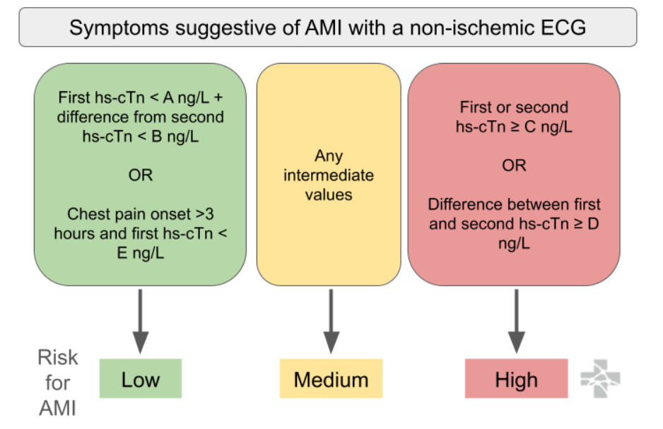
    2. rapid rule-out of ACS with a non-ischemic ECG and either 1) one very low troponin result (depending on onset time of chest pain >3hrs) OR 2) very low change between two consecutive troponins (aka low delta troponin) 

  • Below is an algorithm from Ali-EM for recommended use. Troponins are recommended by the AHA/ACC to be trended every 3 hours, if trending is clinically indicated. Protocols are institution-specific.  

Interpretation of High-Sensitivity Troponin

  • Normal values vary based on type of high sensitivity troponin (institution-specific) and sex: 

    • High Sensitivity Troponin I (hs-TnI) Males: <20 ng/L and Females: <15 ng/L 

    • High Sensitivity Troponin T (hs-TnT) Males: <14 ng/L and Females: <9 ng/L

  • Limitations 

    • Because high sensitivity troponin tests detect lower troponin levels, there are more likely to be false positives for ACS detection (especially in chronic illness / stress / stable CAD / HF / CKD). This may result in unnecessary testing and invasive measures. Further clinical trials are required to guide clinical-decision making in these situations. Troponins should only be ordered when clinically relevant, and should be carefully interpreted based on clinical context. 

 ·