
A 32 yo female with PMH of nephrolithiasis and arthritis presenting with a few hours of severe, intermittent, worsening R flank/back pain radiating to the groin, “just like my kidney stones.” One episode of vomiting from the pain. No urinary symptoms. Currently on her menstrual period. Vital signs within normal limits.
POCUS bladder done by Dr. Odashima showed:
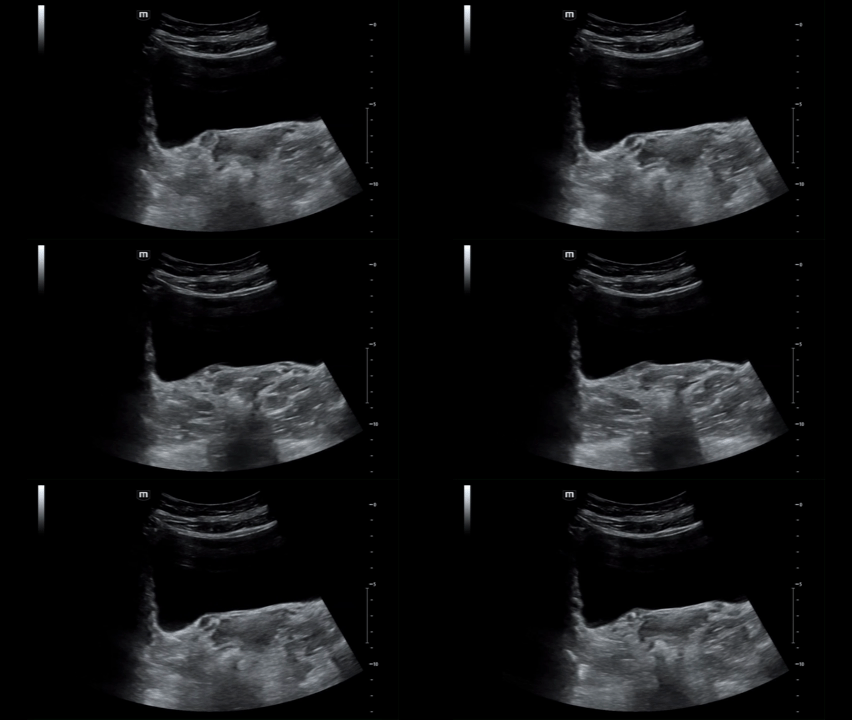
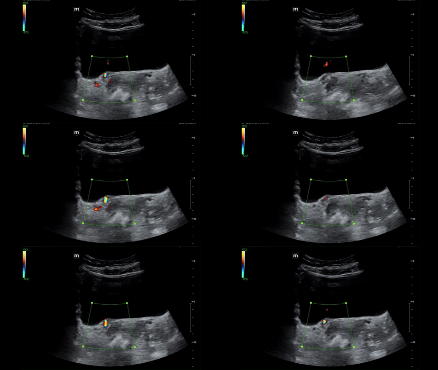
The grayscale image shows a bright, echogenic stone at the UVJ. Then we turn on color Doppler and the stone lights up like a disco ball, a mosaic of rapidly alternating red and blue behind a rough, strongly reflective surface. That is the twinkle artifact: color signal where nothing is actually flowing. It happens because the stone’s rough, irregular surface scatters the ultrasound beam in a random way, and the machine reads that as motion. Twinkle artifact can help you pick out stones that are small or hard to see on grayscale alone.
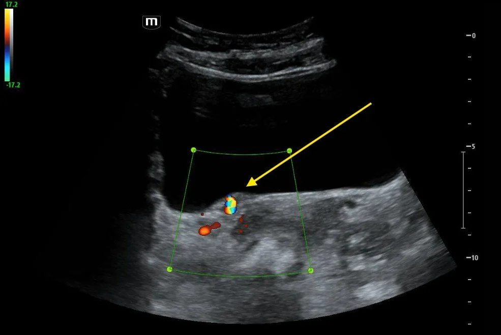
Twinkle artifact
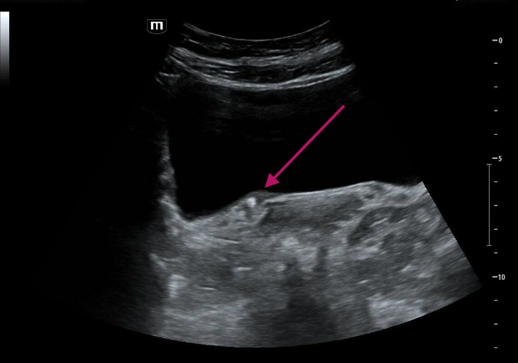
UVJ stone
UA was negative. A dose of ketorolac relieved her pain, and she was discharged with medical expectant management and urology follow-up *high five*
Clinical pearls:
Twinkle is not a diagnosis, but it can guide management. Twinkle has a pooled sensitivity of about 86% and specificity of about 92% for urolithiasis, so it’s a great rule-in tool, but a negative scan doesn’t rule out a stone. Other calcifications, gas, and even renal vessels can mimic it. It’s also sensitive to machine settings, so adjust your color gain and settings if it isn’t showing.
Spare the radiation! Patients with kidney stones often get scanned again and again, and the cumulative CT dose adds up. In patients with a known stone history and a classic presentation, POCUS (stone, twinkle, hydronephrosis) can often answer the question without another CT. Save CT for when you’re worried about something else: fever, infection, a solitary kidney, an unclear diagnosis, or pain that doesn’t fit.
Thanks to Kay for sharing this cool case with us!
-Ultrasound team
References:
Rahmouni A, Bargoin R, Herment A, Bargoin N, Vasile N. Color Doppler twinkling artifact in hyperechoic regions. Radiology. 1996;199:269-271.
Kamaya A, Tuthill T, Rubin JM. Twinkling artifact on color Doppler sonography: dependence on machine parameters and underlying cause. AJR Am J Roentgenol. 2003;180:215-222.
Katz SI, Saluja S, Brink JA, Forman HP. Radiation dose associated with unenhanced CT for suspected renal colic: impact of repetitive studies. AJR Am J Roentgenol. 2006;186(4):1120-1124.
Broder J, Bowen J, Lohr J, Babcock A, Yoon J. Cumulative CT exposures in emergency department patients evaluated for suspected renal colic. J Emerg Med. 2007;33(2):161-168.
Smith-Bindman R, et al. Ultrasonography versus computed tomography for suspected nephrolithiasis. N Engl J Med. 2014;371:1100-1110.
Diagnostic accuracy of Doppler twinkling artifact for identifying urolithiasis: a systematic review and meta-analysis. J Ultrasound. 2023;26:321-331. doi:10.1007/s40477-022-00759-z