
71 yo male with pmh of HTN, DM2 presents with 2 days of painless left-sided vision loss. Pt reports of seeing “only the periphery” out of his left eye, with loss of central vision and the nasal visual field. Pt denies fall or trauma. VS stable. On exam, pt has a noticeable visual deficit in L eye, EOM intact, PERRLA.
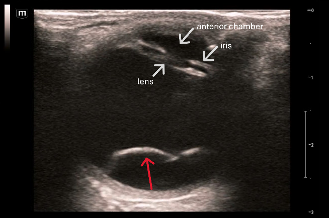
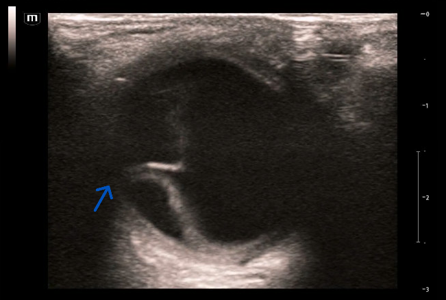
Bedside POCUS showed:
This is an example of a retinal detachment. POCUS has a reported sensitivity up to 96.9% for the diagnosis of retinal detachment. In Figure 1, the red arrow points to a thick hyperechoic membrane representing the detached retina. The retina can be seen lifting off the posterior surface of the globe and moving with eye movement. In Figure 2, the retinal detachment is seen on both the temporal and nasal sides of the retina, remaining tethered to the optic disc (blue arrow). This is an important distinction from posterior vitreous detachment (PVD), which appears as a thinner, more mobile line, and not tethered to the optic disc. PVD is typically not an ophthalmic emergency.
Mac-on/Mac-off:
Mac-on: the macula remains attached
Mac-off: the detachment has extended to involve the macula, resulting in central vision loss.
Because the macula is located temporal to the optic disc, a detachment involving only the nasal retina may be mac-on and require immediate surgery.
Given this patient's significant central visual deficit and sonographic findings, this was concerning for a macula-off retinal detachment. This patient was sent to the retina clinic at Kings County the same day for definitive management.