
4yo female with no significant pmh presents with left hip pain. When she woke up this morning to get ready for daycare, pt was unable to walk due to L hip pain. Mom denies any trauma or recent falls, denies any fevers. On arrival, vital signs are stable, afebrile. Exam shows no significant redness or swelling to the L hip. Pt has left-sided antalgic gait and ROM of L hip limited by pain. Xrays of bilateral hips, L femur, and L tib/fib are unremarkable.
Bedside POCUS revealed:
You can see here anechoic fluid in the anterior synovial space between the femoral neck and posterior surface of the iliopsoas muscle. This is characteristic of a hip effusion. The sonographic criteria for diagnosing a hip effusion is fluid measuring > 5 mm or > 2 mm of fluid difference between the affected and the contralateral normal hip. These measurements are made from the anterior bony cortical surface to the posterior surface of the iliopsoas muscle at the concavity of the femoral neck [red line in figure above] (Tsung et al., 2007).
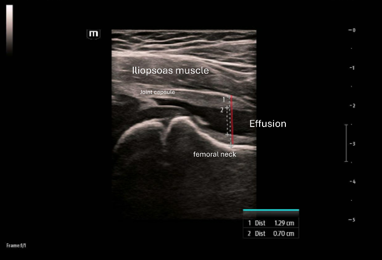
Clinical Pearl: Seeing a hip effusion on POCUS alone cannot differentiate between septic arthritis and benign causes, such as transient synovitis. Clinical picture is very important (are they febrile, toxic appearing, can they bear weight, etc.), and in many cases further workup may be needed. This patient’s bloodwork showed no leukocytosis, ESR and CRP were WNL. Pt tested positive for rhino/enterovirus and was discharged with strict return precautions. Pt’s mom was called to follow up a few days later, pt is now walking normally and no longer has any pain.