
An 85 year old female with PMH of HTN, CHF, DM, NSTEMI, hypothyroidism presented with painless vision loss in the left eye. Symptoms started 1.5 hours prior to presentation. In the ED, patient states that her vision is blurry and can only see out of a "horizontal slit" in her left eye.
Vital signs were stable. Exam showed L eye complete visual field defect. pupils equal, L eye sluggish but reactive, EOM intact.
Stroke code was called, and initial CT imaging showed no acute intracranial pathology. Bedside POCUS showed the following finding:
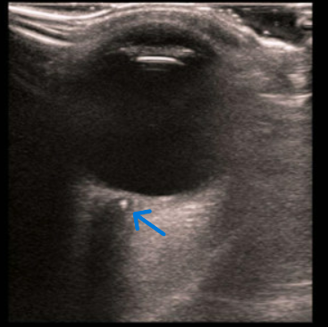
Color Doppler can also help distinguish CRAO as there would be diminished central retinal arterial flow on Doppler. Normally, you should be able to see normal arterial and venous flow detected posterior to the globe.
The patient was taken for emergent MRI/MRA to assess for concurrent acute stroke, which was negative in this case. Stroke team recommended dual antiplatelet therapy, and patient was admitted for further workup, including ophthalmology evaluation, carotid US, and Echo.
Clinical Pearl: CRAO is considered an ophthalmic emergency with a high risk of long-term vision loss due to retinal ischemia. CRAO most commonly occurs from carotid artery atherosclerosis and cardiac embolism (evaluate for afib), but giant cell arteritis is a possible cause of CRAO and must be ruled out.