
A 54 year old male with pmh of atrial fibrillation came in with palpitations. HR was in 140s and BP 113/85. EKG showed atrial fibrillation with RVR.
Bedside POCUS showed a large pericardial effusion:
Classic signs of cardiac tamponade were seen:
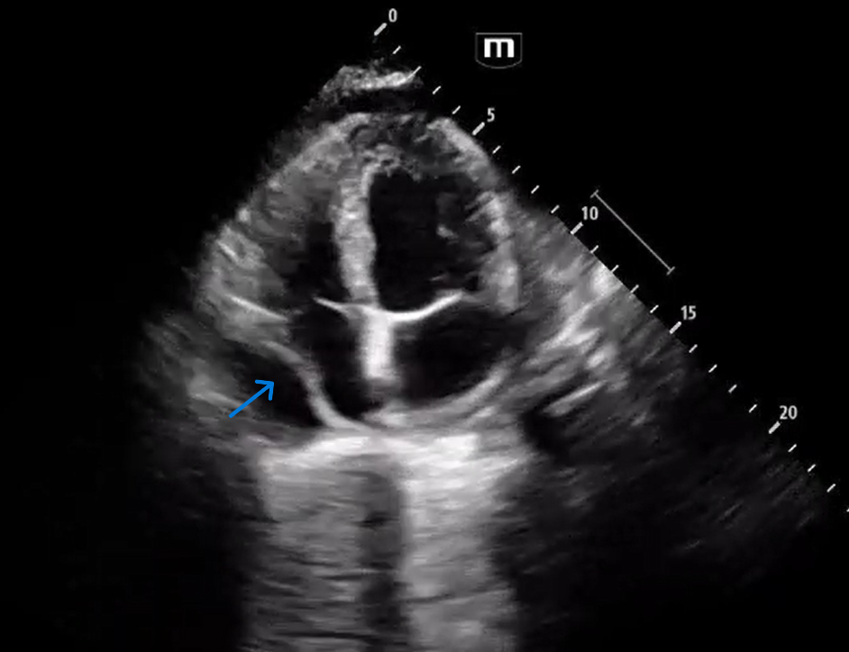
A4C shows circumferential pericardial effusion, RA systolic collapse and RV diastolic collapse.
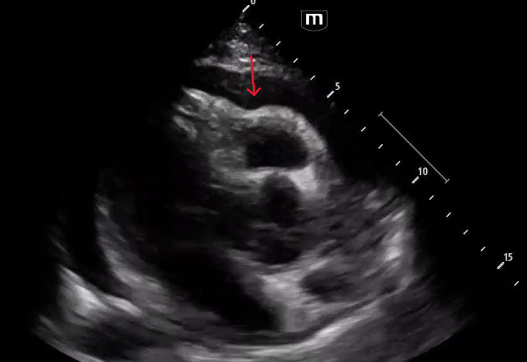
PSS shows effusion and RV collapse and sonographic alternance: "beat-to-beat" variation in the swinging or rocking motion of the heart.
PSL shows RV diastolic collapse
Plethoric IVC
blue arrow shows RA systolic collapse:
red arrow shows RV diastolic collapse:
Course: Patient was resuscitated with iv fluids, and CT surgery was immediately consulted. Patient’s heart rate improved to 80s and patient’s blood pressure remained stable.
Cardiology echo confirmed tamponade physiology, and the patient was taken urgently to the OR for pericardial window with pericardial drain placement and was admitted to the SICU for further monitoring and management. CT also showed a left lower lung mass and bilateral pleural effusions, and the pericardial fluid cytology is positive for non-small lung carcinoma, demonstrating that this was a malignant pericardial effusion due to metastases.
Key Points:
The rate of fluid accumulation is the most critical factor in determining whether a pericardial effusion leads to cardiac tamponade, causing obstructive shock. The pericardium requires time to stretch and accommodate larger fluid volumes.
Right atrial collapse during systole is a highly sensitive and specific sign for cardiac tamponade. It is one of the earliest signs of cardiac tamponade physiology (right atrium is not as muscular as the RV and is the first to be affected against high intrapericardial pressure)
Right ventricular collapse during diastole is a more specific and usually later sign for cardiac tamponade.
You can use M mode on the ultrasound machine to determine RV collapse in diastole in PSL view:
M-mode- displays motion of structures along a single scan line (white dotted line above) over time.
Diastole occurs between the red dots where the mitral valve opens for LV filling. The yellow dot represents the pericardium. The blue dot represents the epicardial border of the RV collapsing during diastole.
Resuscitation Tip: fluid resuscitation is key, especially as a temporizing measure before pericardiocentesis. The heart is preload-dependent while it tries to overcome the overwhelming intrapericardial pressure. Vasopressors, like Norepinephrine, can be used as well if the patient remains hypotensive. However, definitive management is an emergent pericardiocentesis in an unstable patient.