High Sensitivity Troponin

Today I will be discussing the high sensitivity troponin test for diagnosing acute coronary syndrome in the emergency department. Many emergency departments nationwide (and worldwide) have transitioned towards using high-sensitivity troponins. It is crucial to understand the test, benefits compared to standard troponin, use, and interpretation. 


High Sensitivity Troponin vs. Standard Troponin for Detection of ACS 

  • High sensitivity troponin recommended by the American College of Cardiology (ACC) in 2022 as gold standard for use in diagnosing ACS in the emergency department. Plethora of evidence demonstrating high sensitivity troponin's ability to detect:

    • A greater number of MIs within 30 days (without change in mortality rate)  

    • Lower concentrations of troponin compared to standard troponin (mild/subclinical injury)  

    • ACS earlier in course, often 1-3 hours after myocardial injury


Use of High Sensitivity Troponin in the ED 

  • Recommended use in patients with symptoms concerning for ACS in the ED: 

    1. Obtain high-sensitivity troponin

    2. rapid rule-out of ACS with a non-ischemic ECG and either 1) one very low troponin result (depending on onset time of chest pain >3hrs) OR 2) very low change between two consecutive troponins (aka low delta troponin) 

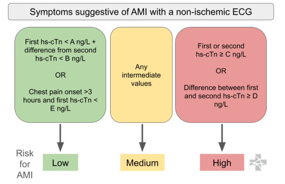
  • Below is an algorithm from Ali-EM for recommended use. Troponins are recommended by the AHA/ACC to be trended every 3 hours, if trending is clinically indicated. Protocols are institution-specific.  

Interpretation of High-Sensitivity Troponin

  • Normal values vary based on type of high sensitivity troponin (institution-specific) and sex: 

    • High Sensitivity Troponin I (hs-TnI) Males: <20 ng/L and Females: <15 ng/L 

    • High Sensitivity Troponin T (hs-TnT) Males: <14 ng/L and Females: <9 ng/L

  • Limitations 

    • Because high sensitivity troponin tests detect lower troponin levels, there are more likely to be false positives for ACS detection (especially in chronic illness / stress / stable CAD / HF / CKD). This may result in unnecessary testing and invasive measures. Further clinical trials are required to guide clinical-decision making in these situations. Troponins should only be ordered when clinically relevant, and should be carefully interpreted based on clinical context. 

 · 

VOTW: What did the lung say to the liver? We be-lung together!

Hello all! This week’s VOTW is brought to you by yours truly!

Hospital course

8 y/o M with PMH asthma presented after 5 days of URI-like symptoms with worsening shortness of breath yesterday. On exam the patient was tachycardic and had active subcostal retractions and crackles on auscultation. A bedside thoracic ultrasound was done.

Ultrasound

Shown above is the right hemidiaphragm or right upper quadrant view. We can see the liver and lung with the diaphragm (dotted line) in between. Note that the lung looks very similar in appearance to the liver! This liver-like appearance of the lung is called ‘hepatization’.

In the image above the lung contains small, dark linear structures that are called air bronchograms. They represent air-filled bronchi.

Case Conclusion

The patient continued to have respiratory distress and developed status asthmaticus requiring PPV. CXR showed bilateral atelectasis and possible underlying airspace disease concerning for pneumonia. The patient was admitted to the PICU on HFNC and given asthma treatments and IV antibiotics.  

Lung hepatization & air bronchograms

·       This patient had a combination of both lung atelectasis and pneumonia. Typically, lung tissue is not visible on ultrasound considering that it is normally filled with air. However, in the case of pneumonia, alveoli are filled with inflammatory fluid creating consolidations, while in the case of atelectasis the alveoli are collapsed rather than fluid filled. This pathologic lung tissue is now visible with ultrasound!

·       Lung consolidations change the appearance of lung tissue on ultrasound such that its echogenicity looks remarkably liver-like, termed ‘hepatization’. This can also be seen in atelectasis.

·       Air bronchograms visible on ultrasound represent air trapped within the small bronchi which are surrounded by lung tissue. Static air bronchograms are mostly seen in compression atelectasis but can also be seen in pneumonia; they are hyperechoic air-filled bronchi that do not move with respirations. Dynamic air bronchograms are pathognomonic for pneumonia; they represent air bubbles moving through fluid filled lung tissue. On ultrasound, dynamic air bronchograms look like numerous hyperechoic opacities that move with respirations.


Happy scanning!                                                                              

Sono team


Resources to review:

·       https://www.acep.org/sonoguide/basic/lung

·       https://coreultrasound.com/5msblog-dab/

·       https://www.thepocusatlas.com/lung/

 · 

VOTW: Hip, Hip hooray!

Hello all! This week’s VOTW is brought to you by myself!

Hospital course

70 y/o F presented to the ED after a fall onto her left hip. XR imaging confirmed a left femoral neck fracture. Instead of using IV opioids, a PENG block was done to relieve the patient’s pain!

Ultrasound

Above we can see the important anatomical landmarks of the pericapsular nerve group (PENG) block. The femoral artery and veins (FA, FV) were identified using color doppler as shown. The bony land marks are the anterior inferior iliac spine (AIIS) and iliopubic eminence (IPE), with the ilium in between. The psoas tendon (PT) is seen along the groove created by the iliopsoas notch.

Here we can see the needle inserted under the psoas tendon with injection of local anesthetic which hydrodissects, or lifts, the psoas tendon off the bone. The spread of the local anesthetic is indicated by the dotted blue line.  

See the clips to see the needle insertion and hydrodissection in action!

Case Conclusion

The patient had excellent pain relief after the PENG block and the patient was taken to the OR later that day for surgical repair.

PENG (Pericapsular Nerve Group Block) block

·       This block targets the terminal sensory branches of the femoral, obturator, and accessory obturator nerves. It is ideal for pain control in the setting of intertrochanteric hip and femoral neck fractures, as well as acetabulum and pubic rami fractures. This block primarily targets sensory nerves, preserving motor function!

·       The probe should first be placed in transverse orientation over the proximal thigh, inferior to the inguinal ligament. After identifying the femoral head, the probe should be moved up until the AIIS and IPE of the ilium are visualized. Key anatomical landmarks to note are the femoral artery and psoas tendon which runs along the groove created by the iliopsoas notch. In general, the femoral nerve lies above the psoas tendon, lateral to the femoral artery.

·       The needle should be inserted in a lateral-to-medial approach until it contacts the ilium bone underneath the PT. Hydrodissection of local anesthetic should lift the PT off the ileum.

·       When inserting the needle take care to avoid going near the femoral nerve and femoral artery!


Happy scanning!                                                                              

Sono team


Resources to review:

·       https://nerveblock.app/nerve-blocks/peng/

·       https://www.acep.org/emultrasound/newsroom/apr2021/pericapsular-nerve-group-peng-block-for-patients-with-hip-or-pelvis-fractures-in-the-ed

·       https://www.nysora.com/education-news/the-hip-block-new-addition-to-nysoras-web-app/

·       https://www.acepnow.com/article/benefits-of-using-the-pericapsular-nerve-group-peng-block/

 ·