VOTW: A Tale of Two Lumens

58 y/o male with PMHx of HTN, CVA in 2013, seizures, and 40+ PPD smoking hx was transferred from outside hospital for aortic dissection. Patient reports worsening back, abdominal pain, nausea x2 days with associated bilateral lower extremity pain, weakness, and inability to walk.


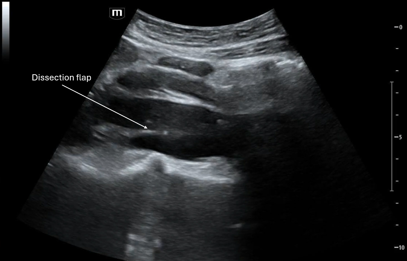
Clip 1 shows a transverse view of the proximal aortal with a dissection flap between what appears to be the true lumen and the false lumen.

As the clip goes on we see the branch points of the celiac trunk and the SMA, it appears that the dissection involves the celiac artery.

In Clip 2, the longitudinal view of the aorta includes the celiac trunk and the SMA, again it appears that there is a flap in the celiac trunk but not in the SMA.

Clip 3 is another longitudinal view of the aorta with the dissection flap.

CTA Chest/Abdomen/Pelvis revealed a type B dissection from the aortic arch to bilateral external iliacs, with a dissection flap extending into the celiac artery with SMA and R renal artery originating from the true lumen.

POCUS Pearls for Aortic Dissection

  • Scan the aorta in chest/back/abd pain with shock or syncope

  • Abdominal aorta should be < 3 cm, Aortic root should be < 4 cm

  • Look for dilation, flap, double lumen, and pericardial effusion

  • Obtain both transverse and longitudinal abdominal views of the aorta; parasternal long axis can visualize the beginning of the aortic root with the suprasternal notch view to visualize the aortic arch

  • Can utilize color doppler to distinguish true lumen (faster flow) from false lumen (slow/swirling)

  • Red flags: aortic root dilation + pericardial effusion → Type A dissection until proven otherwise

  • Negative POCUS ≠ rule out aortic dissection, CTA if clinical suspicion is high

  • If you see anything abnormal → escalate immediately

References:

  1. Nazerian P et al. POCUS protocol for acute aortic syndromes. Acad Emerg Med. 2019.

  2. Moore CL et al. US in aortic emergencies. Acad Emerg Med. 2007.

  3. Evangelista A et al. Key imaging findings in acute aortic dissection. Circulation. 2003.

  4. Kimura BJ. Focused echo for aortic root dilation. JASE. 2012.

  5. Nienaber CA & Clough RE. Acute aortic syndromes overview. Lancet. 2020.

 · 

VOTW: Not So FAST

This week's VOTW was brought to you by Dr. Emily Cen and Dr. Akshara Ramakrishnan!

42 y/o male with PMHx of gastric perforation (s/p repair 10+ years ago) presents to the ED sent in by PMD for abdominal pain in setting of fall of 1.5 meters at construction site 4 days ago with +FAST at PMD's office. Patient states that he went to his doctor today because of right upper quadrant abdominal pain and 1 episode of vomiting yesterday. Denies lightheadedness, dizziness, chest pain, shortness of breath.

The team did a FAST and here are some labeled images from their respective clips:

Clip 1 is a LUQ that shows free fluid below the diaphragm on top of the spleen. 

Clip 2 shows a RUQ view with the liver that appears to have a heterogenous mass.

Clip 3 shows a transverse pelvic view with free fluid in the abdomen, likely blood with a mix of fresh and coagulated blood based on the different echogenicity. Not to be confused with the bladder that is sitting right under all that free fluid and the bowel gas with dirty shadowing over to the right.

Clip 4 shows a sagittal pelvic view with bowel over to the left with dirty shadowing, again seeing the large volume of free fluid in the pelvis with the bladder off to the right of the screen. 

From the imaging, patient may have liver cancer, and the trauma from the fall seemed to have caused the masses to bleed and cause large volume hemoperitoneum. IR wasn't able to identify the exact vessel that had been bleeding but empirically embolized the right hepatic artery. A total of 2 liters of bloody fluid was drained from the abdomen.

Here are some general reminders about the FAST exam:

  • RUQ view is most sensitive – Look at diaphragm → Morison’s → inferior kidney pole with fluid often first at liver tip

  • LUQ is more posterior + superior than RUQ – Look at splenorenal, subdiaphragmatic, inferior pole

  • Pelvis views with both sagittal + transverse – Look posterior to bladder (rectovesical)

  • Cardiac view can be switched to parasternal long if subxiphoid is poor – Look at pericardial vs pleural: pleural fluid lies behind descending aorta

  • Free fluid is anechoic with sharp angles between structures but can appear complex if clotted

  • FAST finds fluid, not source

  • Possible false negatives with early shock, retroperitoneal bleed, body habitus

References:

Kirkpatrick AW et al. Focused Assessment with Sonography for Trauma (FAST) Consensus. J Trauma. 2020.

Stengel D et al. Accuracy of FAST Meta-analysis. BMJ. 2001.

American College of Surgeons (ACS) ATLS 10th Edition.

Wish you all happy scanning!


 · 

Principles of ED Fracture Reduction

Hello Everyone, 

Today I’ll be discussing the principles of closed fracture reduction in the emergency department. At MMC Main, we are lucky enough to have a responsive orthopedic surgery team available to us- however, this may not be the case out in the community. Knowing the basic principles of fracture reduction is crucial to restoring orthopedic function, particularly when neurovascular compromise is present. 

General Indications for Fracture Reduction by the ED: 

Closed fracture

- If open: consult orthopedics and administer TDAP and antibiotics

Displaced fracture

- If non-displaced or minimally displaced, then proceed with a splint- reduction is not needed 

- If angulated, comminuted, intraarticular, delayed 

presentation, or physeal in children: consult orthopedics for possible surgical 

fixation 

Location of fracture 

-ED fracture reductions are only indicated for certain anatomical locations. Be sure to verify whether orthopedic management is mandatory for the injury at hand. 

Quick plug for a mobile application called “Fractures.app” (EMRA-endorsed and a cousin of “sutures.app” and “nerveblock.app”) that provides indications and techniques for reduction, splinting, and follow up recommendations based on anatomical location of fracture. 

Preparing for Fracture Reduction: 

1. Establish mechanism and neurovascular status

2. Obtain X-rays to confirm fracture

3. Pain control: Consider IV pain medications, hematoma block, procedural sedation, nerve block, intraarticular block

Hematoma Blocks can be a low-risk and efficient pain control method: 

  1. Obtain 5-15cc of 1% lidocaine in a syringe (toxic dose of lidocaine without epinephrine is 5mg/kg) 

  2. Identify the fracture site and clean with antiseptic solution 

  3. Create a wheal of anesthetic at the area superficially above fracture site 

  4. Insert the needle tip into the wheal until the bone is felt while aspirating - aspiration of blood indicates correct placement in the fracture hematoma (make sure it is not pulsatile in the syringe or continually flowing to avoid systemic injection) 

  5. Inject lidocaine 

Basic Principles of Fracture Reduction: 

1. Longitudinal traction-countertraction 

2. Recreate and exaggerate mechanism of injury while holding traction-countertraction 

3. Alignment of the fractured ends while holding traction-countertraction. Then release traction to stabilize.  

4. Post-reduction XR to verify alignment 

4. Immobilization via Splinting/Casting

6. Reestablish neurovascular status 

Here is a link to the full 3-minute video demonstrating a hematoma block, distal radius fracture reduction technique, and splinting: 

https://youtu.be/cy6f7he2e4w?si=SVXqBZvoqmVVyAds

Make sure to provide the patient with orthopedic follow up, splint care instructions and return precautions as indicated.

Best,

Lekha Reddy 

 ·