VOTW: Eye for an Eye

40 y/o male with no PMHx presents for sudden visual changes in the right eye starting yesterday morning with blurry vision in the inferior visual field of the right eye. States that he was seeing "white bubbles,” and a “wave” that is black with some red discoloration.  Reports that the vision changes worsened acutely with the "dark area" appearing to expand upward. Denies eye pain, trauma, or prior similar episodes. States that he wears glasses at baseline with no other ocular diseases.

In Clip 1 there is a thick bright hyperechoic line that extends from the optic disc, very much tethered to the back of the eye ball, that is the retina that has detached from the back of the eye.

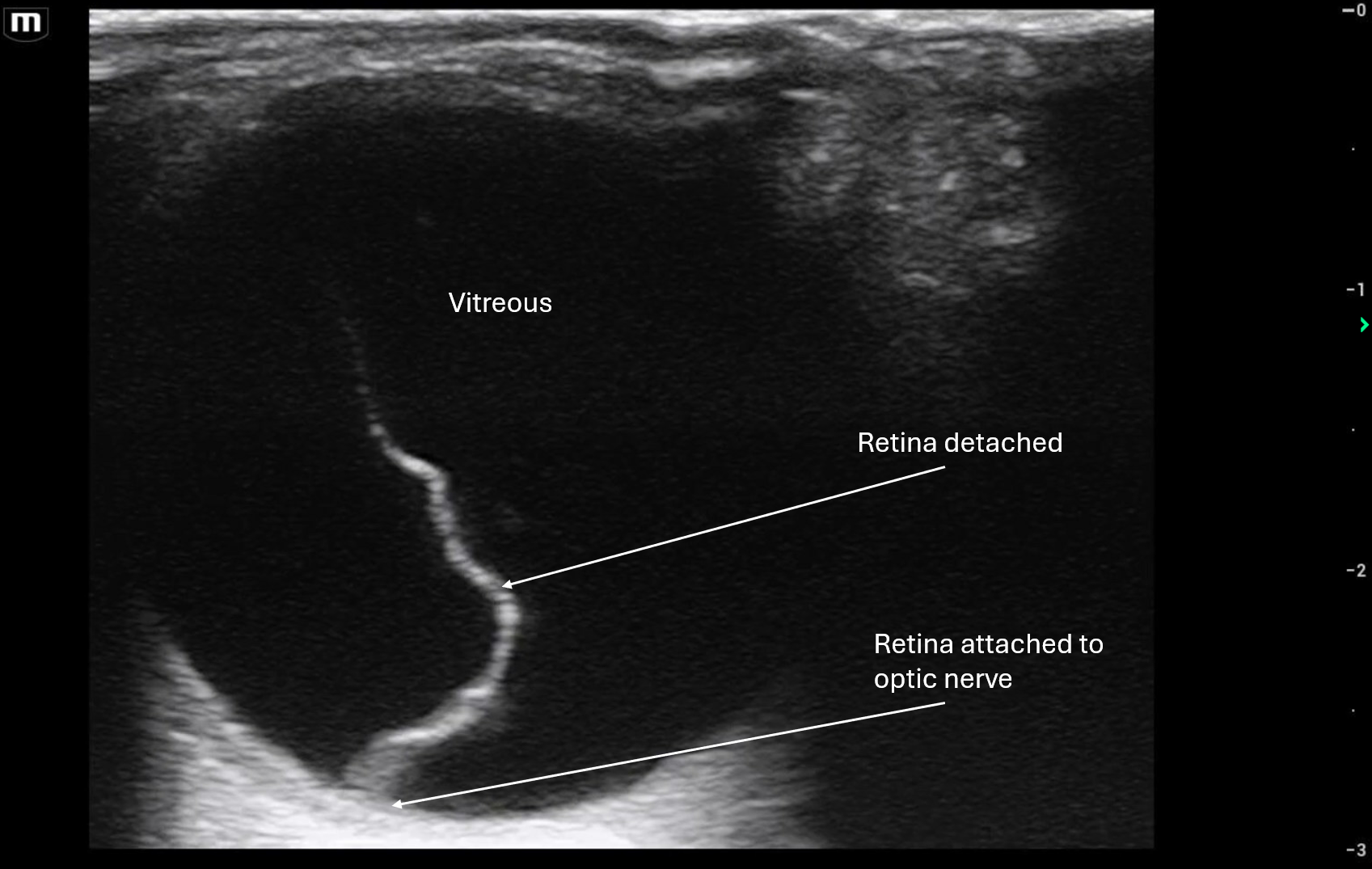
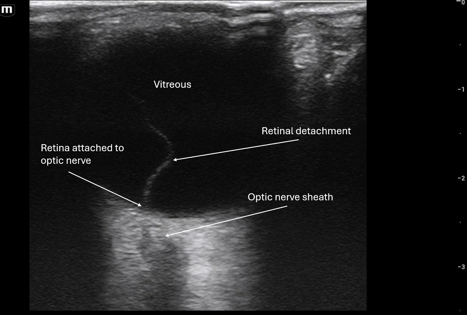
In Clip 2 it shows that the retina does seem attached to the optic disc because you can see the optic nerve sheath in this image.

In both clips you can see that when the patient moves his eye, the retina is mobile but attached to the optic disc/back of the eye. This is concerning for retinal detachment until proven otherwise!

Patient was transferred to outside hospital to Ophthalmology for retinal detachment.

POCUS Pearls for Retinal Detachment Ocular Ultrasound:

  • Hyperechoic, thick linear membrane within the vitreous

  • Tethered to the optic disc (key distinguishing feature)

  • V-shaped, Y-shaped, or funnel-shaped configuration in larger detachments

  • Limited mobility with eye movement (moves less than vitreous debris)

  • Often described as a “sail” or “curtain” floating in the posterior chamber

 · 

VOTW: Blood or Bowel?

76 y/o male with no reported PMHx presented via EMS for acute SOB after fall at home. Per EMS, neighbors called EMS after hearing fall as patient lives alone, reportedly admitted recently for a fall in Oct 2025 and declined nursing home upon discharge. EMS found patient to be hypoxic to 70% on RA and improved on CPAP. Patient placed on BiPAP by team with good O2 saturation but decompensated after having an episode of coffee ground emesis on BiPAP with hypotension and abdominal pain. Patient found with diffuse abdominal tenderness and bedside POCUS of the abdomen was completed.

Video Block
Double-click here to add a video by URL or embed code. Learn more

In all 3 clips we see the right upper quadrant, and in different cuts of it you can see that there is some sort of complex fluid surrounding the liver and between the liver and kidney. It isn't anechoic or hypoechoic like we would expect of free fluid like ascites or even blood from an acute bleed, it could be coagulated blood but usually that has more of a layering effect than swirling of hyperechoic and hypoechoic contents.

It may also appear that there is some free fluid around the bowel loops and possibly some bowel wall edema which all points to perforated bowel and resulting global bowel inflammation.

Patient was unstable and due to episode of coffee ground emesis was presumed to be GI bleed with perforated viscus. Patient was taken emergently to the OR without a CT scan and they found "large volume feculent peritonitis, global intraabdominal necrosis (SB, colon, stomach, liver, diaphragm, peritoneum), 1cm cecal perforation." Patient's prognosis was grave and died several hours after the OR.

We are very sorry for his family's loss and respectfully thank him for this learning opportunity.

POCUS Pearls

  • Free blood

    • Anechoic to hypoechoic when acute

    • Becomes heterogeneous / mildly echogenic as it clots

    • Usually smooth, homogeneous, without internal architecture

  • Stool / bowel contents

    • Heterogeneous, mixed echogenicity

    • Often dirty shadowing

    • May have mottled, granular appearance

    • Can show gas with reverberation or ring-down appearance

 · 

POTD: Burn

Today we'll go in the opposite direction of frostbite and talk about burns. While Maimonides is not a burn center, we still do get burn patients coming in. 

Classifying Burns:

We no longer use 1st, 2nd or 3rd degree but based this off the depth of the burn

Superficial thickness: involves the epidermal layer only

Skin looks red

Superficial partial thickness:  involves the superficial dermis. 

Can cause blisters, looks wet, more painful

Deep partial thickness: involves the deep dermis

Looks cherry red or pale, now causing delayed cap refill

might be less painful now

Full thickness: Involves all the layers including fascia, muscle, bone

looks white, waxy, leathery. No cap refill

usually painless in that area

Burns can worsen over time and the full extent may not be known immediately

Estimating Burn size:

Only include partial and full thickness used to estimate seize. 

Rule of 9's and the Palmar Method are two ways to estimate the size of the burn


Wound Care:

Run wound under cold water if possible and you can use soap to clean it. Avoid ice or toothpaste as this may worsen the injury.

Keep the wound moist to aid in healing and use a non-adherent pad 

Silver Sulfadiazine used to be recommended but may actually slow healing so no longer recommended. Bacitracin can be used for superficial wounds. 

Blisters: sloughed, necrotic or ruptured blisters can be debrided but otherwise small unruptured blisters should be left alone. 

Airway Considerations:

Keep a high suspicion for impending airway compromise. There may be inhalation injury. 

Signs for early intubation include: respiratory distress, stridor, hoarseness, burns to the face and neck, blisters in the oral mucosa

You can also consider using a scope to visualize patient's supraglottic area to see if there is edema, erythema

Indications for ED Escharotomy:

Escharotomies in burn patients are considered when the injury is extensive enough to cause compartment syndrome

Indications include: 

Inability to ventilate patient due to burns to the torso, absent or decreasing pulses, neuro deficits

Escharotomy is done to the burn areas with eschar and not to the fascial layers like a fasciotomy


Fluid Resuscitation:

These patients have impaired skin barriers and are likely to be dehydrated and it is important to fluid resuscitate them

Parkland Formula is widely used to calculate the amount of fluids to give to these patients. However there is concern of causing fluid overload and edema. 

The Modified Brooke's can be used and this allows for less initial fluids to be given to hopefully prevent fluid overload. 

Infection: 

These patients are at higher risk for infection. However, the use of prophylactic antibiotics is controversial. 

Antibiotics should be given when there is proven infection or the patient is septic. 

Indications for transfer:

Full thickness burns

Partial thickness and >/= 10% TBSA

Partial or full thickness involving hands/feet, face, genitals, over joints

Burns with other traumatic injuries

Uncontrolled Pain

Inhalation injury

Chemical Injuries

High Voltage Injuries (>1000)

Lightning Injury