Hello all! Check out this week’s VOTW by yours truly!
Hospital course
A 60 y/o M with extensive PMH including ESRD on dialysis and CHF presented to the ED complaining of generalized weakness and SOB. He was hypotensive and anemic. Bedside TTE was performed.
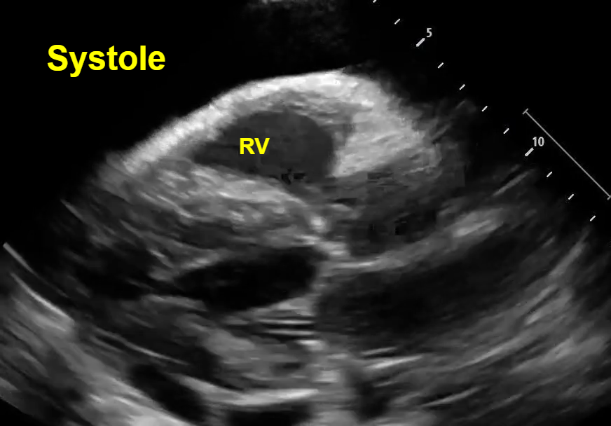
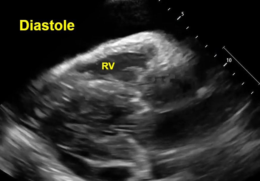
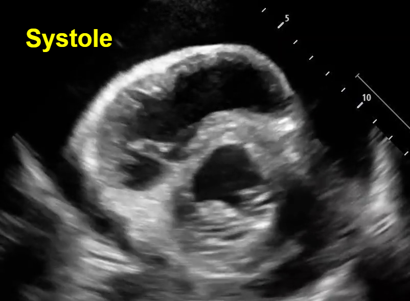
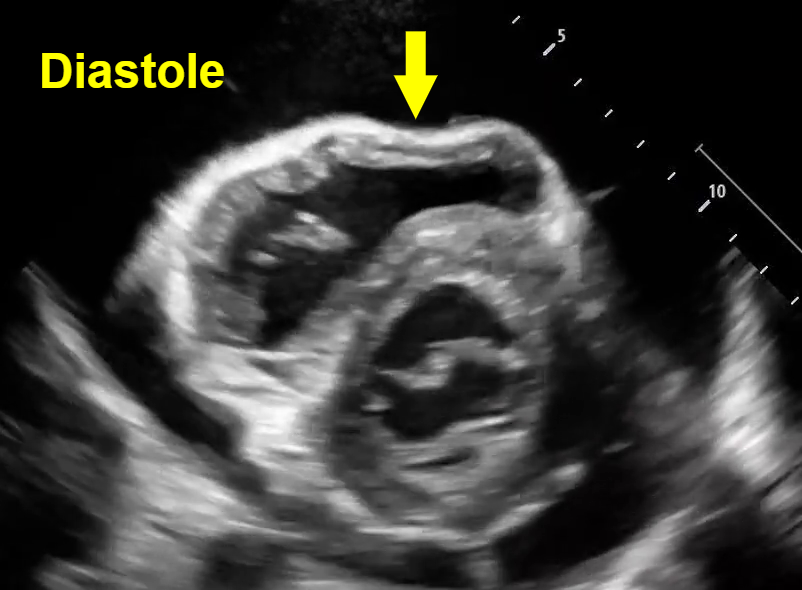
In both parasternal and short axis views seen above, there is a circumferential pericardial effusion surrounding the entire heart.
Parasternal long view: We can see the RV collapse during diastole. How do we know this is diastole? Note that we can see the opening of both the mitral valve and the tricuspid valve to allow for ventricular filling, which occurs during the diastolic phase of cardiac contraction. See clip #1 to see a video of this RV diastolic collapse.
Short axis view: Here we see an example of ‘trampoline sign’, which is the characteristic bouncing motion of the RV. In the image above, we see inversion of the RV wall during diastole (arrow). How do we know this is diastole? Again, note that we can see the opening of the mitral valve in the LV when the RV wall inverts. See clip #2 to see a video of the ‘trampoline sign’.
IVC: In clip #3, we see a very distended plethoric IVC without respiratory variation.
Case Conclusion
The patient was found to have a large pericardial effusion with tamponade. He was stabilized and admitted to cardiology for a pericardial window.
Characteristic Findings of Cardiac Tamponade on POCUS
· The transition from a pericardial effusion to tamponade is due to the rate of fluid accumulation within the pericardial sac, not the total volume of effusion. The right heart is a low-pressure system and collapses when it is unable to accommodate the acute increase in pressure seen when fluid quickly fills the surrounding pericardial sac.
· Thus, the earliest sonographic finding of cardiac tamponade is RA collapse during systole. This is typically followed by RV collapse during diastole, which has both high sensitivity and specificity for cardiac tamponade.
· A non-collapsible plethoric IVC is the most sensitive sign of cardiac tamponade.
Happy scanning!
Sono team
Resources to review:
· https://coreultrasound.com/pericardial-tamponade/
· https://www.acep.org/emultrasound/newsroom/may-2024/cardiac-tamponade
· https://www.aliem.com/differentiating-pericardial-effusion-tamponade-ultrasound/
· https://www.emra.org/emresident/article/us-cardiac-tamponade