VOTW: Let’s put a knee-dle in that knee!

Hello all! This week’s VOTW is brought to you by myself.

Hospital course

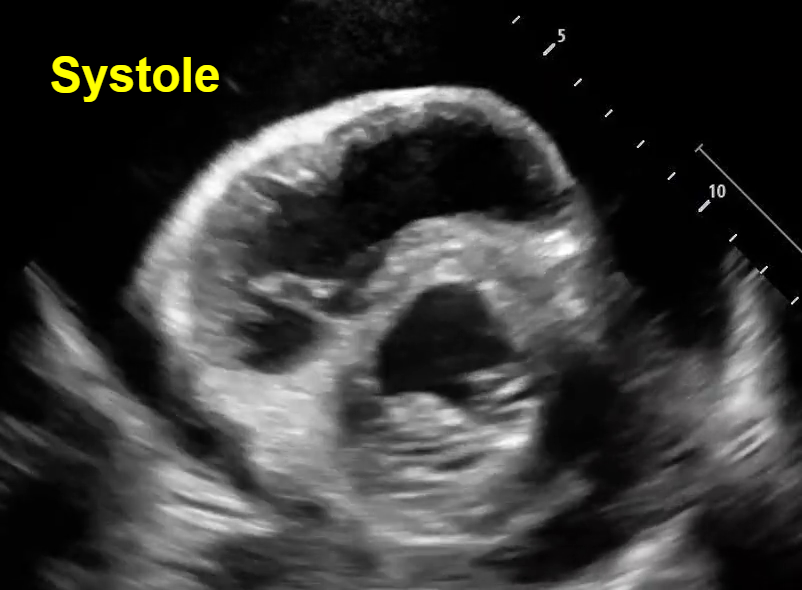
50 y/o F presents to the ED with several weeks of lower back and left knee pain. Left knee is swollen and tender with palpation. She limps when ambulating due to the pain. Bedside ultrasound of the knee joint is shown below.

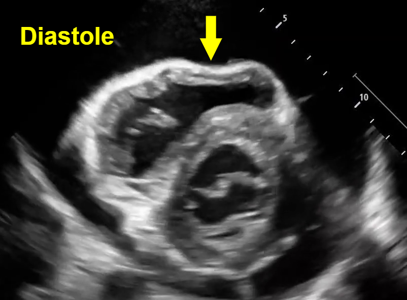
In the image above, the probe is placed over the suprapatellar space with a long-axis view. The femur is seen below, with a layer of dark anechoic effusion visible between the quadriceps tendon and the prefemoral fat pad.

Once the suprapatellar effusion was identified, the probe was rotated 90° into a transverse view of the knee, and the needle was inserted lateral to medial using an in-place approach. In the image above you can see the needle tip enter the effusion underneath the quadriceps tendon. The needle is the hyperechoic straight line in the left image, which is labelled with a white arrow in the right image.

In the clip above we can see this ultrasound-guided in-plane needle insertion with the needle tip entering the suprapatellar effusion.

In this clip, we can see the effusion shrink in size as the synovial fluid is actively aspirated through the needle!

Case Conclusion

The synovial fluid specimen was sent to the lab, and septic arthritis was ruled out. The patient’s left knee pain significantly improved after the arthrocentesis and she was discharged.

Ultrasound-guided knee arthrocentesis

·       While this procedure can be performed blind, the use of ultrasound improves accuracy, improves pain scores, and allows aspiration of more synovial fluid.

·       The patient should be positioned supine with the knee in 15-20° of flexion.

·       Begin by identifying a suprapatellar effusion by placing the linear probe superior to the patella with the marker oriented cephalad (long axis view of the knee). A joint effusion will look like an anechoic stripe within the joint space deep to the quadriceps tendon.

·       Once you identify a drainable effusion, rotate the probe 90° to obtain a transverse view (short axis view of the knee). A lateral to medial in-plane technique should be used to guide the needle tip into the joint effusion.

·       Remember to use a sterile ultrasound probe cover!

·       Ultrasound-guided arthrocentesis can be used to drain effusions from any joint, with commonly aspirated joints including knees, hips, shoulders, wrists, elbows, and ankles.

Happy scanning!

Sono team

Resources to review:

·       https://www.acep.org/sonoguide/procedures/arthrocentesis

·       https://mskultrasound.net/arthrocentesis-of-the-knee/

·       https://coreultrasound.com/knee-aspiration-and-injection/

 · 

VOTW: A real gut-wrenching situation!

Hello all! This week’s VOTW is brought to you by yours truly!

Hospital course

83 y/o F with PMH esophageal hernia presented to the ED with 2 days of abdominal fullness, nausea, and vomiting. Last bowel movement was 2 days ago. Bedside ultrasound was done.

This is small bowel. How do we know this? Note the small finger-like projections from the inner wall (yellow arrows). These are called plicae circulares, which are  mucosal folds of the small intestine. Also note that the bowel diameter is dilated up to 3.2 cm (blue arrow).

Note the transverse view of the small bowel below the stomach. The bowel wall appears thicker than normal, measuring 0.88 cm. Also note that the stomach itself appears very dilated!

In the clip above, we can see multiple loops of dilated small bowel. We can see hyperechoic specs of intestinal contents within the bowel making a “to-and-fro” motion instead of normal unidirectional peristalsis.

In the clip above, we can see dilated small bowel with no movement of the intestinal contents at all!

Case Conclusion

The patient was found to have a small bowel obstruction with an incarcerated femoral hernia on CT imaging. NG tube was placed in the ED and patient was admitted for surgical intervention.

Characteristic Findings of SBO

·       In normal small bowel, the regular bowel diameter is < 2.5 cm and we expect to see normal peristalsis with unidirectional flow.

·       A small bowel obstruction on ultrasound will show multiple loops of bowel with a diameter > 2.5 cm. The intestinal contents will appear to move back and forth with “to-and-fro” movement. Sometimes the distal obstruction will prevent the intestinal contents from moving at all!

·       A small bowel obstruction will also lead to bowel wall edema, which causes a wall thickness > 4 mm. The bowel wall edema and dilated diameter will make the intestinal folds or plicae circulares appear more prominent, leading to the “key board” sign seen in the image above.


Happy scanning!

Sono team


Resources to review:

·       https://coreultrasound.com/small-bowel-obstruction/

·       https://www.emdocs.net/us-probe-ultrasound-for-small-bowel-obstruction/

·       https://www.acep.org/sonoguide/advanced/gi---bowel-obstruction

 · 

VOTW: The chamber that bounced back!

Hello all! Check out this week’s VOTW by yours truly!

Hospital course

A 60 y/o M with extensive PMH including ESRD on dialysis and CHF presented to the ED complaining of generalized weakness and SOB. He was hypotensive and anemic. Bedside TTE was performed.

In both parasternal and short axis views seen above, there is a circumferential pericardial effusion surrounding the entire heart.

Parasternal long view: We can see the RV collapse during diastole. How do we know this is diastole? Note that we can see the opening of both the mitral valve and the tricuspid valve to allow for ventricular filling, which occurs during the diastolic phase of cardiac contraction. See clip #1 to see a video of this RV diastolic collapse.

Short axis view: Here we see an example of ‘trampoline sign’, which is the characteristic bouncing motion of the RV. In the image above, we see inversion of the RV wall during diastole (arrow). How do we know this is diastole? Again, note that we can see the opening of the mitral valve in the LV when the RV wall inverts. See clip #2 to see a video of the ‘trampoline sign’.

IVC: In clip #3, we see a very distended plethoric IVC without respiratory variation.

Case Conclusion

The patient was found to have a large pericardial effusion with tamponade. He was stabilized and admitted to cardiology for a pericardial window.

Characteristic Findings of Cardiac Tamponade on POCUS

·       The transition from a pericardial effusion to tamponade is due to the rate of fluid accumulation within the pericardial sac, not the total volume of effusion. The right heart is a low-pressure system and collapses when it is unable to accommodate the acute increase in pressure seen when fluid quickly fills the surrounding pericardial sac.  

·       Thus, the earliest sonographic finding of cardiac tamponade is RA collapse during systole. This is typically followed by RV collapse during diastole, which has both high sensitivity and specificity for cardiac tamponade.

·       A non-collapsible plethoric IVC is the most sensitive sign of cardiac tamponade.


Happy scanning!

Sono team


Resources to review:

·       https://coreultrasound.com/pericardial-tamponade/

·       https://www.acep.org/emultrasound/newsroom/may-2024/cardiac-tamponade

·       https://www.aliem.com/differentiating-pericardial-effusion-tamponade-ultrasound/

·       https://www.emra.org/emresident/article/us-cardiac-tamponade

 ·